| 9 |
The malaria of mangrove swamps and Anopheles ludlowi |
| 9.1 |
The Philippine islands |
“Being in Manila early in 1901,” wrote Dr. Clara S. Ludlow, B.Sc, Ph.D. [12,13], “it was repeatedly suggested by some of the medical officers of the United States army stationed there that I should take up the study of mosquitoes, and the thought strongly emphasised that the study of mosquitoes was likely to be of real benefit to mankind, especially if carried out in connection with the occurrence of certain diseases, notably malaria.” It was a period when the mosquitoes of tropical countries were being studied in detail for the first time. Miss Ludlow, who had accompanied her brother, a colonel in the army, to Manila, was a student of science, well qualified for the task; and the work was regarded as so important that it was put under the jurisdiction of the Surgeon-General. Thus began a research, which was continued for many years in the Philippines, and is still carried on by Dr. Ludlow at the Army Medical Museum, Washington, where I had the pleasure of meeting her in 1913.
Dr. Ludlow soon found [12] that “… the mosquitoes taken were not described in the books available, and in a little while it became evident that no one knew the mosquitoes of the Philippine Islands.” She worked, however, in conjunction with Mr. Theobald of the British Museum, and rapid progress was made in describing the species taken. At quite an early date (1903) she differentiated the three members of the rossi group of Anopheles, although for many years to follow this interesting and important group remained a puzzle to most workers. The group consists of three mosquitoes, A. rossi, Giles, A. indefinita and A. ludlowi; the interest lies in the fact that they are so much alike that for long many workers did not regard them as separate species: the importance, in that only A. ludlowi is an important natural carrier of malaria, while the innocence of the other two has been generally accepted. Another point of interest is that A. ludlowi breeds mainly along the coast and in brackish water. Our knowledge of these mosquitoes has come slowly, and even now is probably incomplete.
| 9.2 |
Anopheline species |
Anopheles rossi (type Giles), the first member of the group to be recognised, was described by Giles from specimens taken in India. It was studied by the Commissioners sent to India by the Royal Society; and their conclusion was that, although it could be infected with malaria experimentally, it played practically no part in propagating the disease in nature. They showed there were wide areas where it existed in which the spleen index was low; if found in malarious places, there was also present another Anopheles species, in which alone malaria parasites were to be found.
Anopheles indefinita is common in the Malay Peninsula. It differs from A. rossi (Giles) in minor details. The evidence is against it being a carrier of malaria, for it is regularly found in large quantity where the spleen rate is low. Its larvae are to be found in small muddy pools and puddles, especially when polluted by the presence of animals or man.
Anopheles ludlowi.—The story has been more involved. Dr. Ludlow found it to be the species of Anopheles most frequently taken in the Philippines, and it was associated with malaria in such a way as to give her the impression that it was an important carrier of malaria—although this was not actually proved by dissection. She noted that it was found, not only along the coast, but also in inland valleys, far from brackish water.
| 9.3 |
Dr. De Vogel’s work |
In Java in 1902 Dr. De Vogel [14,15] found an Anopheles species breeding in salt marshes, often in large numbers in the water of sun-cracked mangrove mud. He noted the prevalence of this Anopheles along the coast, and the existence of high spleen rates:
… 60-100% in the native villages (kampongs) situated along the coast, and in the two overflow canals which carry brackish water far into the land and contain innumerable pools between their banks. Among the rice fields and fresh water marshes farther in the interior, the numbers vary between 5% and 25%.
Dr. De Vogel also experimented with this mosquito and was able to infect it easily; but he was unable to keep the insects alive long enough to trace the parasites to the salivary glands. Specimens were sent to Mr. Theobald, who declared them to be A. rossi, but there can be little doubt, in the light of our present knowledge, that Dr. De Vogel was working really with A. ludlowi. That Mr. Theobald should have made the mistake shows how close the resemblance between A. rossi and A. ludlowi is. To distinguish the two is at times difficult indeed; for in dried specimens the leg spots of A. ludlowi may have faded, and in some A. rossi there are stripes not unlike spots. Mr. R. M. M. Mangkoewinoto [16] has drawn attention to the value of the wing spots when the leg markings are doubtful.
| 9.4 |
Malaria in the Andamans |
It was not until 1912 that the question was practically settled by Christophers [17] in the Andaman Islands, where the convict settlement has been notoriously malarious. On investigation he found that
… malaria in the settlement is confined to a belt around the margins of the harbour, and is absent or nearly so from villages more than half a mile from the sea-coast or the salt swamps associated with this. This freedom from malaria is seen even in inland villages situated on the margins of swamps amidst rice fields and near jungle.
He ascribed this peculiar distribution of malaria to the “distribution of Anopheles ludlowi,” and he found malaria developing in the stomach wall of two mosquitoes out of fifty-three dissected.
It is of interest that A. umbrosus was not found in the Andamans. In 1913 Dr. De Vogel [18], investigating malaria at Sibolga in Sumatra, found it associated with A. ludlowi and described how the spleen rate decreases the farther the kampong is in from the sea.
Finally in 1919 Dr. N. H. Swellengrebel and others [19,20] published an exhaustive study of the association of A. ludlowi with malaria. It is printed in Dutch and English in parallel columns, and so is available for those who cannot read it in the original. The authors are to be congratulated on their investigation, which is a model of what a scientific inquiry should be; and the moderation of their conclusions is altogether admirable. It should be read by every student of malaria.
They found A. ludlowi to be the most important carrier of malaria in the parts of Java and Sumatra in which they worked. They not only infected it experimentally, but also determined its natural index of infectibility for successive months; in each set of observations A. ludlowi is compared with other species of Anopheles. They compare the malarial infection of man and mosquito; and show that, although A. ludlowi is usually confined to the brackish waters of the coast, nevertheless it is found in some fish ponds in inland valleys in which there is alga; that in mangrove forests regularly covered by the tides it does not occur; while it breeds freely in the portions submerged only during spring tides. A picture of sun-cracked mud, which is almost a replica of that published by De Vogel years before, is given.
Now Java and Sumatra are volcanic islands, and the conclusions of these authors support the observations originally made by Dr. Ludlow working in the Philippine Islands—which are also volcanic. In the Malay Peninsula, which is not volcanic, A. ludlowi has never been found away from the coast by either Strickland, Hacker, or myself; and other species are responsible for the inland malaria. When visiting Sumatra in 1913 I was struck with certain differences between that island and the Malay Peninsula; in particular the freedom from malaria of some of their opened hill land. These differences, and the importance of studying the species of Anopheles of a country, I discussed with Dr. Swellengrebel and Dr. Schüffner; and it was this discussion, the authors say, which induced them to begin their research. The impressions I then gathered have been confirmed, and there can be no doubt that important differences exist between the two countries, although they are separated by only a narrow strait.
In towns and villages on the sea-coast or mangrove belt of the Malay Peninsula which I have examined, malaria produced by A. ludlowi alone is rare. A. ludlowi is usually found in association with A. umbrosus; and as good drainage clears away the two insects and the malaria produced by them, the prevention of malaria has in practice presented little or no difficulty in these places [9].
| 9.5 |
Mangrove swamps: two tidal zones |
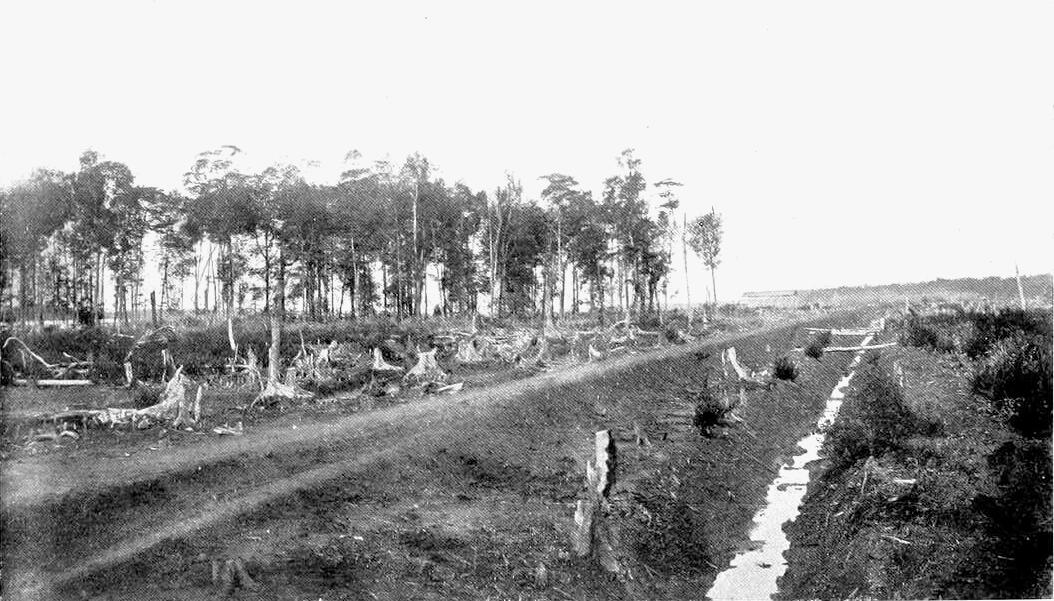
The mangrove forest consists of a number of species of trees which can live only in salt or brackish water; hence they are not found away from the sea-coast or tidal rivers. The forest can be divided roughly into two zones: one nearest the sea, which is covered twice daily and by every tide; the other, farther inland, covered only by spring tides for a few hours in perhaps three days of every fortnight. When the forest is intact, in other words in virgin mangrove forest, no Anopheles breed in the outer zone; and houses built on the sea edge of the mangrove have the reputation generally of being free from malaria. It is otherwise with the inner zone, even in the virgin mangrove. In this inner zone there are, as I [9] wrote in 1903,
… vast numbers of breeding places among mangrove trees and stumps. Here crabs raise mounds, and between these small pools of water are found. Certain of the pools, situated on the zone of land covered by spring tides only, become as the result of heavy rain comparatively fresh-water pools, and at times I have found these at Port Swettenham to be teeming with the larvae of Anopheles. This I regard as the real danger of a mangrove swamp—and there were large tracts of this at Port Swettenham.
Two Anopheles are found here—namely A. umbrosus and A. albotaeniatus. To put it briefly, the outer zone of the virgin mangrove forest is harmless—because no Anopheles breed there; on the other hand the inner zone is malarious because A. umbrosus and A. albotaeniatus live in it; and the former is a known carrier of malaria. As far as I am aware A. ludlowi has never been found in virgin mangrove; and, as Strickland has pointed out, the larvae appear to require sunshine.
When man destroys the forest, and more particularly when he carries out engineering operations in it, one thing certainly happens: he lets sunlight into the land. Another thing may happen: he may prevent the tide from covering and flushing the outer zone twice a day as it naturally does. A railway line along the foreshore is an example of an engineering work which might create extensive areas of stagnant brackish water in the lower zone. In such pools A. ludlowi may breed in enormous numbers; indeed, I think the largest number of larvae of any species ever seen by me in a breeding place has been A. ludlowi; at times they have appeared like a moving pellicle. A. ludlowi are to be found, too, in the inner zone when the forest has been felled. Yet even in felled mangrove the breeding places of A. ludlowi, albotaeniatus, and umbrosus are not identical. For while A. ludlowi is to be seen in large numbers in stagnant pools or blocked drains free from vegetation, the other two will be taken rarely unless there is grass, sticks, or some cover; and they may readily be missed, for they hide themselves away among the black roots of the mangrove and can be captured only by scraping these with the collecting dish.
| 9.6 |
Malaria due to A. ludlowi |
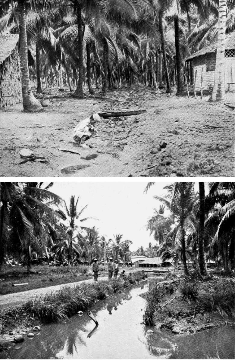
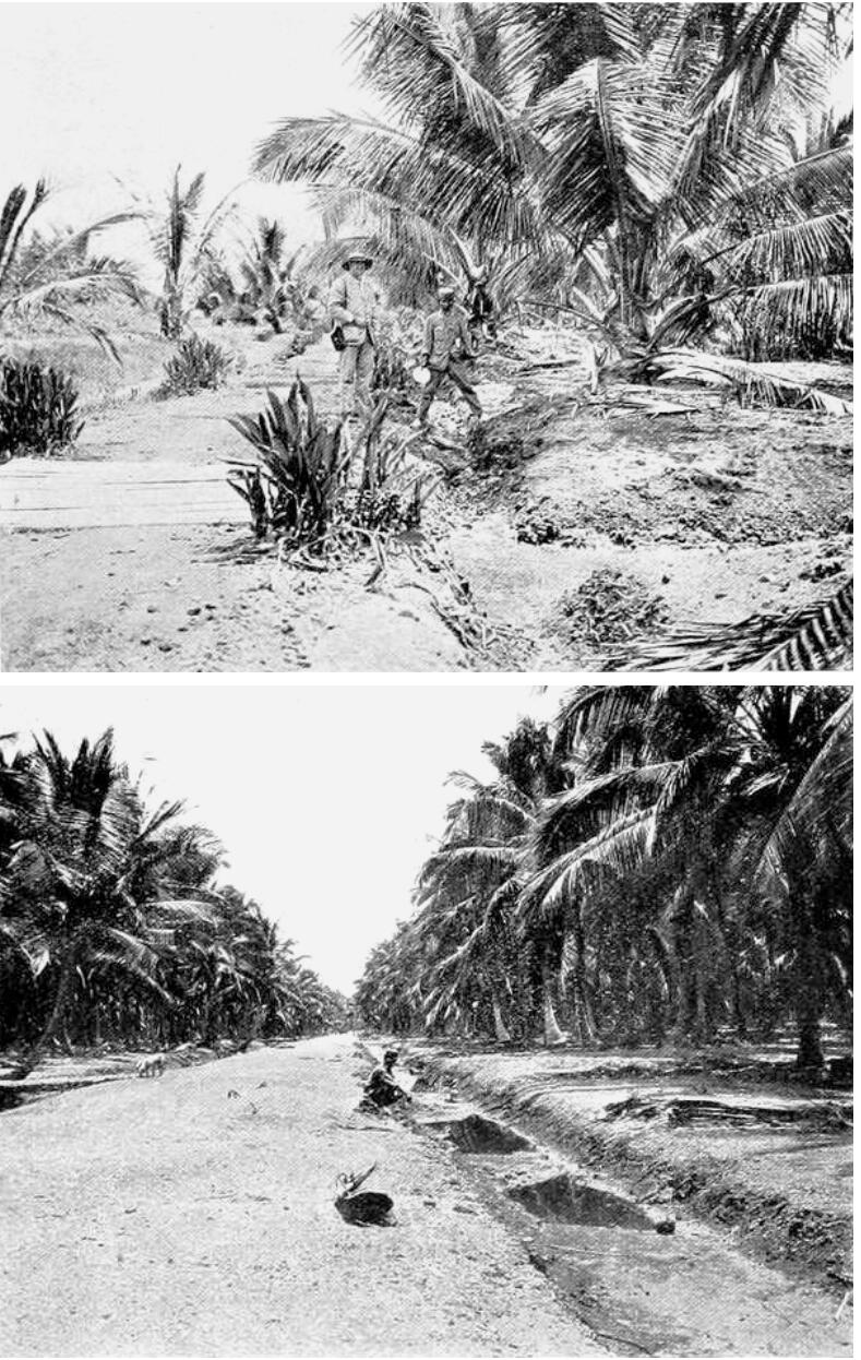
The following is an instance of a place in which I found malaria due apparently to A. ludlowi unaccompanied by A. umbrosus.13 It is a coconut estate near to the mouth of a large tidal river, and is divided into three divisions. The severity of the malaria is indicated from the following spleen rates.
| 9.6.1 |
Division I |
Of 5 Javanese children born on the estate, 4 had enlarged spleens: of 48 Tamil children, 46 (or 95%) had enlargement; of the other two, one had been on the estate four months, and the other one year.
| 9.6.2 |
Division II |
Of 100 children 70 (70%) had enlarged spleen; and of the 30 others only 1 had been on the estate over six months. It is significant that, out of the 100, only 3 were under two years old.
| 9.6.3 |
Division III |
Twenty-three out of 25 (or 95%) children, who had been on the estate over six months, had enlarged spleen. Of 32 under six months on the place—many of them only about one month—no fewer than 14 (or 43%) had enlarged spleen.
A. ludlowi, adult insects, were present in numbers. Immediately on entering my room, I saw two on my mosquito net. In the evening my assistant exposed his arm for about a minute in the open air, was promptly attacked, and captured four.
At daylight on the following morning my assistant searched the twenty-six occupied mosquito nets in the hospital, with the exception of four which were searched by the dresser; my assistant caught Anopheles, the dresser took all mosquitoes present. In only two nets Anopheles were not found. The total catch was 164 mosquitoes, of which 124 were A. ludlowi; the other 40 were Culicines and were almost exclusively Culex sitiens, which, like A. ludlowi, breeds in brackish water.
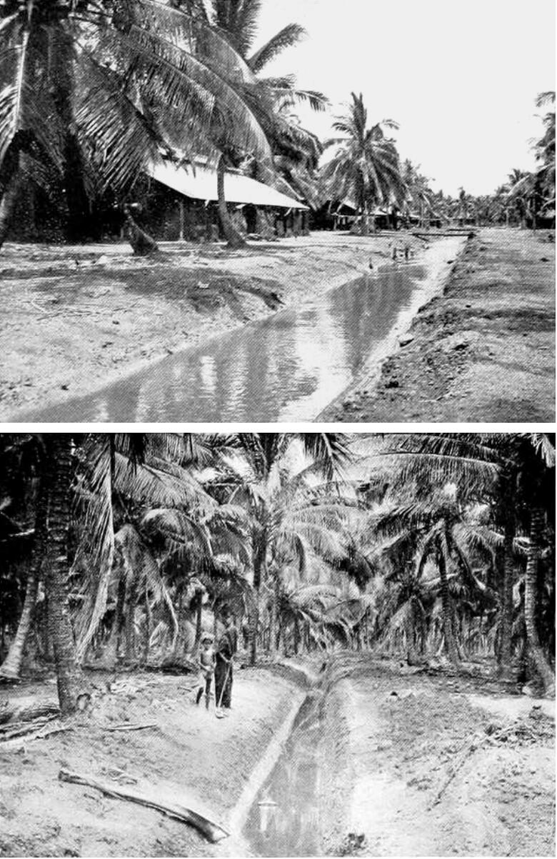
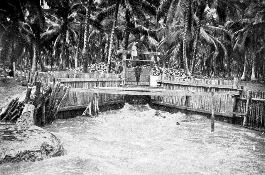
The estate stretches along the river bank, and is protected from tidal flooding by an embankment. At certain times the tide gates are opened to admit sea water into the drains, so that the nuts may be transported cheaply by floating them down the drains. Larvae were found wherever the water was stagnant or vegetation existed. In some shallow drains, free from vegetation, crab holes had formed little land-locked harbours; in these larvae were abundant, particularly so where grass, weeds, or small sticks were present in a pool or stagnant drain. In contrast to these, larvae were not found in the main drains where the edges were free from weeds, clean cut and deep, and where there was current.
| 9.7 |
The control of A. ludlowi on Carey Island |
Since Carey Island became an estate in 1906 it has been under my care. The island is surrounded by large rivers or arms of the sea containing practically pure salt water. The shores of the island were originally covered by the usual Mangrove zone; and as these portions of the land are below high-tide level, the whole is protected by bunds and sluice gates. Beyond the tidal zone the island is flat alluvial soil like the “Coastal Plain” and contains A. umbrosus.
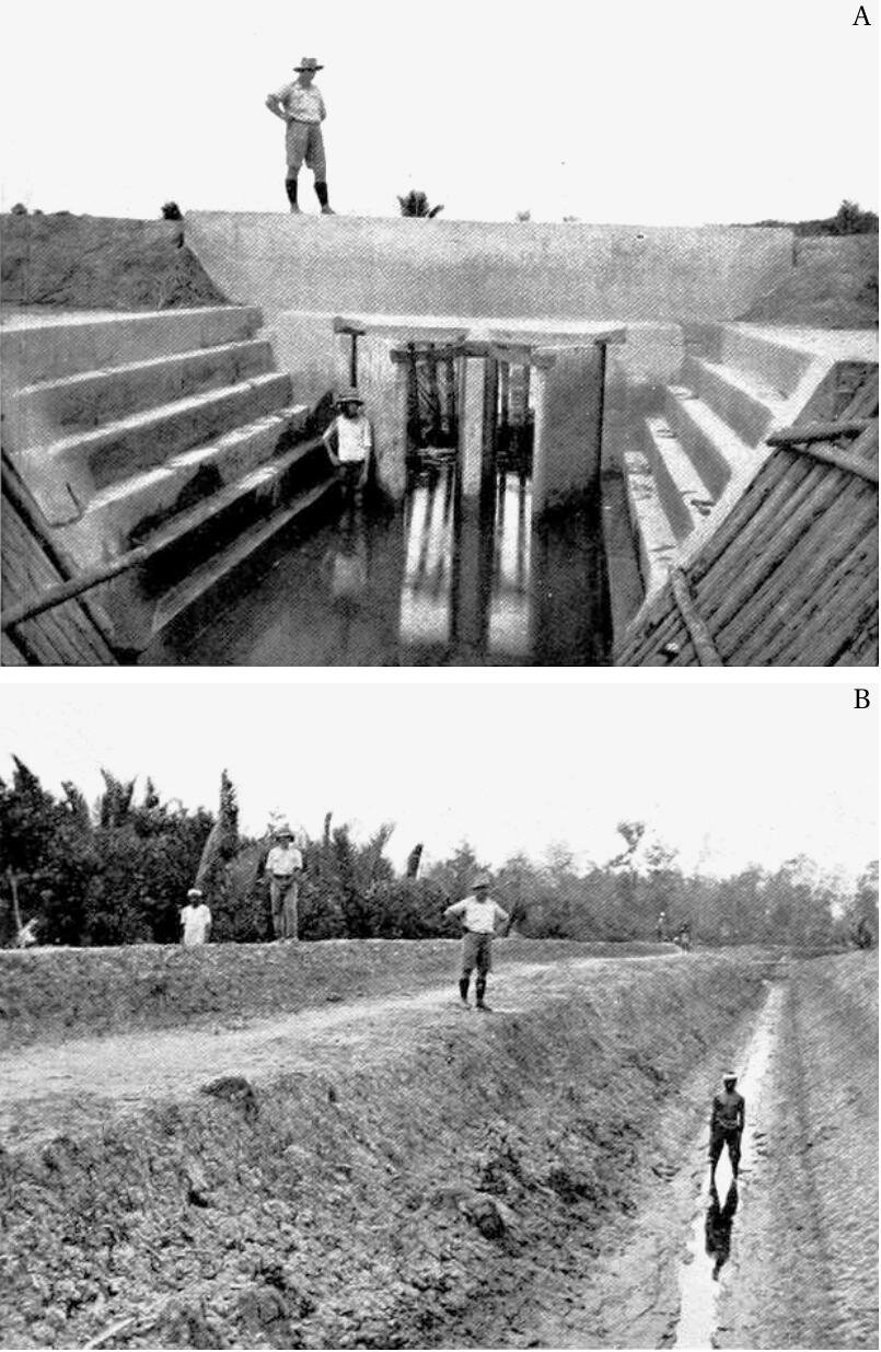
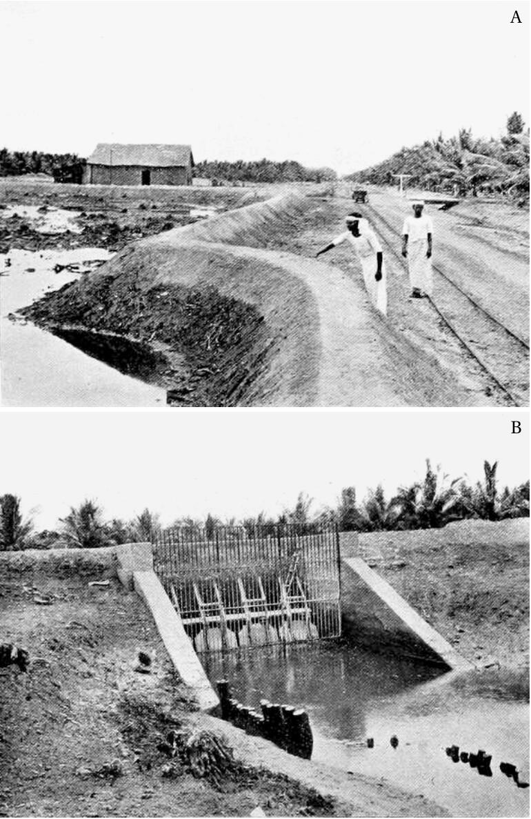
The land and general situation of the island is, indeed, similar to that of the estate previously described. But whereas the estate is and has been intensely malarious, the island has been as consistently free from the disease since the first year it was opened. And the freedom is due to the following simple precautions taken, and the excellent drainage system which exists. On the island full advantage has been taken of the 16-foot rise and fall of the tide; and by means of low-level tide gates, a system of deep drainage has been created far superior to that of the majority of estates on the Coastal Plain of the mainland.
Opening up was begun at a small clearing in the mangrove occupied by Sakais, one of the aboriginal races of the Peninsula. When the island was taken over by the company, 100 acres were reserved for Sakais; but it was bunded and drained by the company; and, until a part of the estate was opened, it formed the company’s headquarters. Drainage, felling, and clearing of the estate land was pushed on rapidly, so that within the first year (1907) 144 acres were planted, and within the second year no fewer than 1828 acres. Each succeeding year saw an increase in the planted area, until by 1917 there were some 10,000 acres opened altogether in two estates.
Now in opening up land, the common, I might almost say, the invariable mistake was to house the labour near to jungle, either temporarily or permanently. This was never done on the island, the definite policy having been adopted, and followed of never housing coolies within half a mile of jungle; and the policy has paid handsomely. In 1909 I wrote of Estate “EE”:
This estate commenced operations at the end of 1906. The labour was at first housed on land originally opened by natives, and drains were at once put through a considerable area by Javanese. So that when the Tamil labour force was introduced later, it was on opened land. The Tamils have accordingly never suffered from malaria, and now one of the finest labour forces in the country is there, consisting in all of about 2000 coolies of whom 1200 are Tamils.
The death rate of the estate was 22‰ in 1908, and in 1909 was only 11.
Owing to the good health of the labour force, over 4000 acres are now drained and opened, and no set of lines is closer than half a mile to the jungle. On 29th December 1909, I examined 246 children on this estate, and found only one with an enlarged spleen (it was just palpable) giving a percentage of 0.4. The boy was in perfect health, and no history of malaria was obtainable. The presumption is, therefore, that he had malaria when in India.
This estate shows that it is not necessarily the oldest estate which is the healthiest; but that old or new the state of the health is in relation to the proximity or otherwise of the jungle.
At the time that was written I was not aware that, by the draining and bunding, we had escaped danger from A. ludlowi in the Mangrove Zone, as well as the danger from A. umbrosus both in the Mangrove Zone and the more central jungle. Hence the reference only to A. umbrosus at that time.
Since 1909 until today, health on the island has been consistently good; indeed, is almost proverbial here. In 1915 on one estate, out of 298 children only 13 or 4.3% had enlarged spleen; and inquiry showed that in almost every case the children with enlarged spleens had been previously employed on estates known to be malarious.
Between 1909 and 1918 the European staff numbered about seventeen, of whom only three suffered from malaria. One contracted his malaria on the mainland, and infected a companion living in his bungalow, which was about 400 yards from jungle. The other, who lived half a mile from jungle, apparently contracted the disease on the island. That was in 1912, and was the last case in the period referred to.
The death rates since 1910 have been 21, 86, 26, 15, 9, 10, 9.12 and 28 in the influenza year, and 5 in 1919, when the average labour force was 2088. The other estate on the island has been equally healthy; the death rates being, since 1912, when it was opened, 0, 10, 17, 13, 22, 14 and 31 in 1918, and 7 in 1919. The spleen rate of 120 children examined in 1915 was 1.6%; one of the two children with enlarged spleen had worked on an estate in Perak, the other in Malacca.
The good health of two estates on the island stands in striking contrast to the malarious coconut estate previously described; and shows how completely the malaria of both the mangrove swamp and Coastal Plain can be controlled by good drainage, and the selection of good sites.
I could give other instances of estates situated on the sea, and within the reach of brackish water, where A. ludlowi and its malaria have been controlled. In Chapter 22, however, I shall trace the recrudescence of malaria in once healthy places on the reappearance of A. ludlowi when breeding places suitable for it have been created. They throw light on the mosquito and malaria from a somewhat different aspect: yet serve to confirm the conclusions already reached.