Author: Michael Palmer

German expat in Canada, MD by training, until recently (March 2022) professor of biochemistry at the University of Waterloo

Human Papilloma Virus (HPV) vaccines do not deserve general recommendation

There is a renewed push underway to inoculate children and adolescents against infections with human papilloma virus (HPV). But is this campaign based on sound science? This post makes the case that there is no urgent need for such a vaccine, and that the existing vaccines have proven neither effective nor safe. On the contrary, the limited available data on the vaccines’ safety raise serious concerns. A general recommendation can therefore not be justified.

Preliminary

This overview relies on a more detailed recent report compiled by Croatian biomedical scientist Dr. Lucija Tomljenovic.1 Her paper also links to more extensive reports composed by herself and by other experienced scientists. All of these documents are freely available online, as is much of the literature which they cite.

The virus

Human papilloma viruses belong to a family of DNA viruses. They primarily infect the skin and the mucous membranes. These infections often manifest as warts. Skin warts are harmless; they may persist for a while but tend to disappear spontaneously when the immune system finally bestirs itself to do something about the situation. Warts on the mucous membranes may affect the genitals, the anus, and the mouth and throat. They are usually transmitted by intimate contact. They, too, will often resolve spontaneously, but in some cases they may not only persist but transform into malignant cancer. This most commonly occurs in the cervix uteri, but the other locations listed above may be affected as well. The likelihood of a progression from mere infection to cancer also depends on the virus subtype. Without vaccination, the subtypes most commonly associated with cancer are HPV-16 and HPV-18. In human populations vaccinated against these two, other subtypes become more prominent.

The purpose of vaccination

The stated goal of HPV vaccination is the prevention of cancer, and in particular cervical carcinoma in women. An early vaccine introduced beginning in 2006 was directed against only the aforementioned subtypes 16 and 18, but in the meantime vaccines directed at up to 9 different subtypes of the virus have come into use. The vaccines are primarily targeted at children and adolescents before they become sexually active.

How effectively does vaccination protect from cancer?

Assessing the efficacy of vaccination against cancer is not trivial, for the following reasons:

  1. the long latency between infection and progression to cancer—even if the vaccination prevented all infections with all HPV subtypes that may cause cancer, a major effect on cancer incidence could only be expected with a delay of several years;
  2. the large potential for bias due to variables such as participation in cancer screening, which in the case of cervical carcinoma is remarkably effective.

Due to the long latency period of cancer, most studies on vaccine efficacy have relied on surrogate markers, i.e. infection as such or cellular abnormalities that do not yet qualify as cancers (“cervical intraepithelial neoplasias”, traditionally called dysplasias). However, it is important to note that early-stage dysplasias may yet spontaneously regress, just like warts.

Among the studies that attempted to assess the prevention of actual invasive cancer, the most optimistic one is that by Lei et al. from 2020.2 These authors evaluated the records, spanning the years 2006 to 2017, of 1.6 million Swedish girls and women between the ages of 10 and 30. They concluded that the incidence of cancer was reduced by 88% in those who had been vaccinated before the age of 17. A more modest effect was reported in another study, which evaluated the results from approximately 350,000 young women from Spain, England and Norway. It found a reduction of dysplasias after vaccination by about 40%, whereas the number of invasive cancer cases was too low for statistical analysis (less than 5 overall).3 While it is not unreasonable to expect that vaccination would reduce the rate of invasive cancer to a similar extent as dysplasias, this is not a foregone conclusion, since detection of a dysplasia by screening would be followed by therapeutic intervention to prevent its progression to actual cancer. In this context, one should note that the optimistic study by Lei et al. did not track the participation of their study subjects in such screening programs. It stands to reason that women who elect to be vaccinated may also be more likely to undergo screening, which could significantly skew the results of the study.

An approach that should not be biased by screening is to compare different types of cervical carcinoma that can be distinguished by histology (i.e. microscopic tissue examination). The two major types are referred to as squamous cell carcinoma and adenocarcinoma, respectively. With both, HPV is found inside the cancer tissue in more than 90% of the cases. However, there remains a “mixed bag” of minor histological types, many of which are not associated with HPV. While it is not easy to determine the exact proportion of HPV-negative cases among these other forms, it probably is less than half. Therefore, if HPV vaccination protected from HPV-induced cancer, than the trends of the major, mostly HPV-positive types and of the minor, often HPV-negative cancers should diverge, such that with time the minor types should account for a greater share of all cervical cancers. This, however, is not observed: while the incidence of cervical cancer among young women has declined very remarkably since 1999, the minor types have declined even faster than the major ones, such that their proportion dropped by about half between 1999 and 2022 (Figure 1).4


Figure 1:
Incidence of cervical carcinoma among women aged 15-29 in the USA, between 1999 and 2022, by histological type. Left: data from Figure S2 in the study by Gopalani et al. Right: fractional contribution of minor histological types to the total number of cervix carcinomas. The trend line is a linear regression.

What might cause the remarkable overall decrease in the incidence of cervical carcinoma? Most likely, it is preventive screening. In keeping with this assumption, other HPV-related cancers—those of the penis, the anus, and the oropharynx—have remained level or even increased over the last several decades. As far as data are available, this also applies to young men, who would be more likely to have received the HPV vaccine than older men. While the limited information does not warrant the firm conclusion that the HPV vaccine has no preventive effect on such cancers, the opposite is equally true—as with cervical carcinoma, the records don’t substantiate any such prevention.

What conclusions should we draw from the limited evidence on cancer prevention?

The lacking evidence of significant protection from cervical carcinoma means that HPV vaccination does not at this time obviate the need for regular cancer screening. Moreover, preventive examination is simple and effective. It is noteworthy that all of the common HPV-related cancers occur at easily accessible body sites. Therefore, just like cervical carcinomas, those of the anus, the oropharynx or the penis could in principle be caught in good time by routine examination. If such preventive exams are deemed unnecessary, then there can be no urgent need for preventive vaccination either.

The HPV vaccines

The HPV vaccines use an interesting technology referred to as “virus-like particles.” These synthetic particles resemble viruses in size and shape, and just like viruses they also carry a large copy number of antigenic protein molecules on their surface. Our immune system tends to respond forcefully to virus particles, and vaccines mimicking their structure likewise tend to elicit a strong immune response. Unlike real viruses, however, these vaccine particles do not contain any copies of the viral genome; they are not to be confused with traditional live vaccines or with the poisonous adenovirus-derived vaccines against COVID-19 that were produced by AstraZeneca and Janssen.

Aside from the highly immunogenic form of antigen presentation, the HPV vaccines also contain a very powerful aluminum-based adjuvant, which serves as a nonspecific trigger of inflammation. This inflammation further amplifies the immune response to the antigen. Overall, therefore, both the antigen preparation and the adjuvant are geared toward eliciting a strong immune response. This might seem like a good thing, but it also poses risks.

The danger of immunological cross reactions

Our immune system wields some powerful weapons—it can attack all kinds of microbes, including bacteria, fungi, parasites, and viruses. To combat viruses, it also destroys cells of our own body which it recognizes as virus-infected. These weapons must of course be deployed with great deliberation in order to avoid harm to ourselves. While overall the immune system’s ability to discriminate “self” from “non-self” is remarkable, it is not infallible. Certain microbes contain antigens that trigger immune responses which attack not only the microbe in question, but also some of the body’s own molecules and cells. A well-known example is the bacterium Streptococcus pyogenes, a common cause of throat or skin infections. These are often self-limiting and also amenable to treatment with penicillin, and in most cases, they heal without any long-term sequelae. However, in some patients, the infection is followed by a severe disease called acute rheumatic fever. In this condition, the immune response that was induced by the infection cross-reacts with self antigens inside the heart, the joints, the kidneys, or even the central nervous system, causing grave harm to these organs. Another example is acute reactive arthritis induced by enteric infections with Campylobacter or Yersinia bacteria; yet another is Guillain-Barré syndrome, a serious disease of the peripheral nervous system, following an infection with the Epstein Barr Virus.

It is important to understand that the risk of such harmful cross-reactions is to a large extent determined by individual genetic traits, and in particular by the constellation of an individual’s histocompatibility antigens. These are the very same genetic markers that also determine the compatibility of organ transplants. You are likely aware that these antigens are highly variable. Just as it is unlikely that I and my next-door neighbor will be a good match for swapping bone marrow transplants, so it is unlikely that we will resemble each other in our susceptibility to autoimmune disease triggered by infectious agents. The same applies to vaccines. It follows that testing a new vaccine on a relatively small collective of volunteers cannot reliably establish the risk of adverse events due to immunological cross-reactions. And with vaccines, there is a further consideration: natural pathogens were present throughout human evolution, so that the most severe adverse responses were removed by natural selection. However, the same does not apply to artificial antigen preparations, i.e. vaccines. Papilloma viruses are of course ancient, too, but a natural infection with such a virus is restricted to one or a few sites on the skin or a mucous membrane, and it induces at best a muted immune response, as is evidenced by the often long persistence of such infections. In contrast, the systemic injection of a large amount of papilloma virus antigen, together with a powerful adjuvant, represents a novel and unusual immunological challenge with unforeseeable risks.5

Can HPV vaccination cause harmful cross-immunity?

Yes, it can. While it is difficult to know exactly how frequent this is, the fact as such has been established by a statistical analysis of the reports collected by the US Vaccine Adverse Event Reporting System (VAERS). To understand the rationale of this analysis, a little background on this system is in order. The purpose of VAERS is to collect putative cases of adverse events that occur after vaccination. While this is useful, there are practical limitations:

  1. not all events that occur are also reported; in fact, the percentage that is reported is likely in the single digits;
  2. some of the events that are reported may not in fact have been caused by the vaccine in question but simply happened close in time due to coincidence.

Therefore, we cannot take the numbers of such reports recorded by VAERS on a given vaccine at face value. However, it stands to reason that the proportion of purely coincidental reports should not depend on the particular vaccine in question. Therefore, if a given vaccine exceeds the average of the other vaccines in the rate of adverse events reported, then at least that surplus of reports can be considered real and substantive.

Dr. Tomljenovic has carried out a thorough comparative study along these lines. She found that certain adverse events were reported significantly more often after HPV vaccination than with all other vaccines combined (with the exclusion of COVID vaccines). Prominent among such events are autoimmune disorders such as systemic lupus erythematosus, juvenile arthritis, multiple sclerosis, and alopecia. The same applies to disruptions of the endocrine and autonomic nervous systems. HPV vaccines are likewise highly overrepresented in reports of embolism and thrombosis. Details can be found in the report cited in footnote 1. These findings are strong evidence of significant potential for immunological mechanisms of harm.

Conclusion

While it is reasonable to expect that HPV vaccines may in the long term reduce the incidence of cancer due to HPV infection, the currently available data to not prove it. The lack of evidence may be due to a multi-year time delay between infection and clinical cancer. However, a long delay also means that there is enough time to catch and avert developing cancers through regular screening. On the other hand, the risk of severe adverse events is real and substantial, even though it cannot be quantified exactly based on the limited data available. Considering that neither the benefit nor the risk can currently be accurately assessed, there simply is no basis for recommending these vaccines to the general public.

Notes

  1. Tomljenovic, L. (2026) Public Comment: Human Papillomavirus Vaccine (HPV) https://archive.org/details/tomljenovic-white-paper (back)
  2. Lei, J. et al. (2020) {HPV} Vaccination and the Risk of Invasive Cervical Cancer. N. Engl. J. Med. 383:1340-1348 http://dx.doi.org/10.1056/NEJMoa1917338 (back)
  3. Alcalde-Herraiz, M. et al. (2026) The effectiveness of {HPV} vaccination against invasive cervical cancer and related precancerous lesions: a multinational target trial emulation study. Lancet Prim. Care 2:100114 http://dx.doi.org/10.1016/j.lanprc.2026.100114 (back)
  4. Gopalani, S.V. et al. (2026) Declines in cervical cancer incidence among young women in the United States. J. Natl. Cancer Inst. 118:941-946 https://www.ncbi.nlm.nih.gov/pubmed/?term=41883186 (back)
  5. One could argue that the most “natural” vaccine yet invented is the polio live virus vaccine, which closely mimics not only the natural pathogen itself but also its route of infection. (back)

The Watson et al. “modeling study”: did “COVID vaccinations” really prevent 14 million deaths?

This piece was written several years ago by a research pharmacist who I believe at this time prefers to remain unnamed.

On June 23, 2022, a “Mathematical Modeling Study” was published in the medical journal THE LANCET Infectious Diseases [1], which purported to show that “COVID vaccinations” have “substantially altered” the course of the pandemic and “prevented 14.4 million … deaths from COVID-19 … during the first year of COVID-19 vaccination.”

Whenever a drug achieves the impossible, it is worth examining the underlying data a little more closely and comparing it to reality.

1. What requirements must normally be met for the approval of a drug or vaccine?

Normally, the benefit provided by a new drug must be demonstrated in extensive clinical studies (“phase 3 trials”) in order for this drug to be approved for the market; this involves a thorough review of the trial documentation by the authorities. For full approval, a pharmaceutical company usually has to submit all of the following:

  • documentation on the drug’s manufacturing quality,
  • preclinical studies (animal studies),
  • phase 1 and phase 2 studies in humans, and
  • 12 months worth of phase 3 trial results that prove beyond doubt the efficacy and the safety of a candidate drug.

The COVID “vaccines” were given “temporary” or “conditional” approval worldwide based on very much shortened phase 3 trials, which lasted only 2 months instead of 12 months [2,3], and on insufficient or missing animal studies, i.e. they received only an “emergency approval.”

The primary clinical endpoints used in the phase 3 studies were not clinically and socially relevant, as predominantly mild, minor events such as headache, cough or fever were counted as “COVID disease cases”, just as long as the RT-PCR test for the coronavirus was positive. An effect of the “vaccinations” on “severe COVID illnesses”, which among other things required hospitalization, was only analyzed secondarily [4,5]. Thus, in order to obtain approval, the manufacturers were not formally required to prove that the “vaccinations” reduce severe courses of disease to a relevant extent.

The fact that the regulatory authorities worldwide accepted this flawed study design for granting approval indicates that they were not able to act independently. To date, the clinical trials have not demonstrated any relevant benefit [6], pivotal documents overall were demonstrably completely inadequate, and moreover the data from the phase 3 studies were manipulated [7–9].

Once a drug is approved, the manufacturer is required to further investigate its efficacy and safety under real-world conditions. The data generated in this way are subsequently submitted to the regulatory authorities, and the results are presented to physicians as “real world evidence” to convince them that these data, ideally, support the findings from the clinical trials and thus to encourage them to use this drug on their patients.

2. What does the reality of “COVID vaccines” show?

The internationally available “Real World Evidence” data of the COVID “vaccines” confirm what the registration studies had already indicated: The “vaccines” are not associated with any relevant benefit, but on the contrary with a negative effect.

no image info
Figure 1: Worldwide daily deaths attributed to COVID-19, March 2020 to December 2021. Graph retrieved from Data [10] on August 24, 2022.

A robust analysis by Kyle Beattie, who examined publicly published COVID data from Our World in Data, shows that for 145 countries, “vaccine” use correlates positively with the number of “COVID cases” and, far more worryingly, with the number of “COVID deaths” [11]. The countries which appear to fare the worst after introduction of the “vaccines” are those where few “COVID deaths” had been reported for 2020 (e.g., Thailand). Based on Beattie’s results, it must be assumed that almost all countries experienced more infections and deaths than if “vaccination” had not been given. Beattie based his model for calculating the hypothetical morbidity and mortality that would have occurred without “vaccination” on the data from four African countries which had very low vaccination rates throughout, and which therefore could be used rather like a “control group.”

Beattie’s conclusions are corroborated by other observations. As of early August 2022, official figures report 6.4 million deaths that have occurred with or from COVID-19. A look at the progression curve of “COVID deaths” recorded worldwide does not reveal any favorable effect on “COVID deaths” concomitant with the roll-out of the vaccines in late 2020/early 2021 (Figure 1). Overall, “COVID deaths” actually increased after the introduction of the “vaccination” and stagnated at a high level throughout 2021. With an effective vaccine, of course, a clearly discernible drop in the mortality curve would have been expected after the onset of a worldwide vaccination campaign.

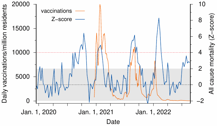
Israel is an instructive example, because it achieved high vaccination rates earlier than most other countries. According to EuroMomo, Israel experienced its highest excess mortality ever since the beginning of the “Corona crisis” in the first quarter of 2022, i.e. at a time when the majority of the population was supposedly maximally protected by the “mRNA vaccines”—even though during this period only the Omicron variant was endemic, which is about ten times less dangerous than the original Wuhan strain and the Delta variant [12]. Increases in excess mortality were correlated in time with vaccination drives (Figure 2). A similar correlation in time is also evident between vaccinations and deaths specifically attributed to COVID-19 (data not shown).

no image info

Figure 2: COVID-19 vaccinations and all-cause mortality (Z-score) for Israel, 2020-2022. The shaded area denotes the normal range for the Z-score; the dotted red line marks the cutoff for a “substantial increase”, according to EuroMomo. Vaccine data retrieved from Data [13], mortality data from EuroMomo [14].

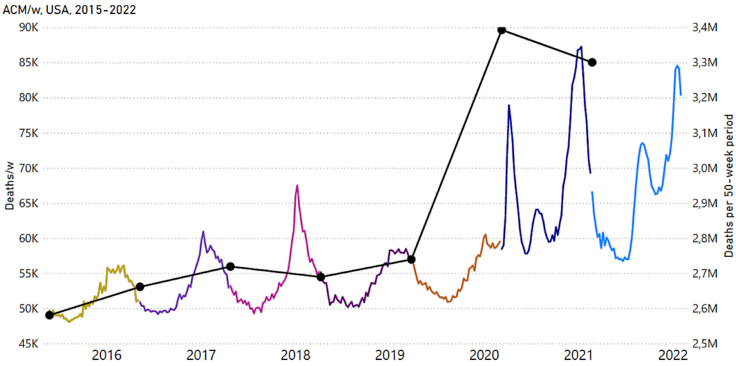
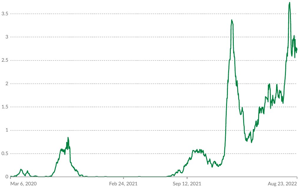
In the USA, the introduction of COVID vaccinations was followed not by a leveling off but by a sustained increase in all-cause mortality (Figure 3). Australia, too, is experiencing a similar phenomenon: After the Australian Department of Health recommended a second “booster dose” of COVID vaccination to all persons aged 30 years and older in July 2022, the country is experiencing a peak in “COVID deaths” of unprecedented proportions, even though the harmless Omicron variant remains predominant (Figure 4).

no image info

Figure 3: All-cause mortality by week (colors) and by 50-week period (black) in the USA from 2015 to 2022. Data are displayed from week-21 of 2015 to week-5 of 2022. The different colors indicate the successive 50-week periods. Adapted from Figure 12 in [15].

3. On what data do Watson et al. base their conclusion that COVID “vaccination” has prevented 14 million deaths?

no image info

Figure 4: Daily confirmed “COVID deaths” per million residents in Australia. Graphic retrieved from Data [10] on August 24, 2022.

The study in question is a mathematical modeling exercise which, like any exercise, depends on the underlying data. A closer look at these data raises serious questions—pertaining not only to the scientific value of this publication, but also to its credibility.

In the “modeling study” by Watson et al., hypothetical data serve as the baseline, not real observations:

“For this mathematical modeling study, we used a previously published COVID-19 transmission model and fitting framework to obtain profiles of the COVID-19 pandemic in each country.”

The authors thus used previously published models to calculate their projections of the hypothetical COVID mortality that would have occurred without vaccination. The problem with these published models is that they date from the early days of the “pandemic” and are based on data and assumptions which were out of date by 2021 at the latest, and which are now demonstrably wrong. In the meantime, it has become clear that SARS-CoV-2 is neither exceptionally dangerous for the general population nor a threat to the health care system. Already at the beginning of the “pandemic”, i.e. at a time when the original Wuhan strain of the virus was prevalent, the infection fatality rate (IFR) was 0.15% overall, less than 0.05% for those under 70 years of age, and 0.00% for children [16]. The IFR of the currently circulating Omicron variant is about 10 times lower than that of the Wuhan strain and the Delta variant [12]. All assumptions on which millions of “COVID deaths” have been estimated worldwide are therefore fundamentally incorrect and obsolete.

4. Watson et al. used inflated “COVID death” rates

It is now known that “COVID deaths” have been massively overcounted worldwide, because both “confirmed cases” and “probable cases” have been lumped together in those case statistics [17]. “Confirmed cases” are counted based on matching (yet non-specific) clinical symptoms together with a positive RT-PCR test result, whereas with “probable” cases no corroborating PCR test result is available. The practice of counting “probable COVID cases” based only on generic symptoms of an upper respiratory tract infection makes no sense from a medical point of view; it can serve only to inflate the case count in an unscientific manner. The Corman-Drosten RT-PCR test protocol for the detection of SARS-CoV-2 [18] is scientifically more than deficient: the test based on it is not validated, not standardized, hypersensitive, and not very specific; its specificity is 98.6% and 92.4% in the absence and presence of other beta-coronaviruses, respectively, and rates of false positive results consequently are 1.4% and 7.6 [19,20].

In only 5% of the “COVID deaths” recorded in the USA was COVID-19 listed as the sole cause of death on the death certificate, and thus an illness due to SARS-CoV-2 was causally responsible for the death [21]. A study from Italy showed that even only 0.8% of the “COVID deaths” did not have a concomitant disease [22]. Instead of taking these facts into account and correcting the case numbers downwards accordingly, Watson et al. even argue that a massive under-reporting of “COVID deaths” must be assumed.

If the enormously high COVID mortality predicted by the modeling were true, then this should have invariably been reflected in a relevant increase in excess mortality during the winter months when the “corona pandemic” was rampant, but prior to the introduction of vaccination.

5. No relevant excess mortality in the 2020 pandemic winter

no image info

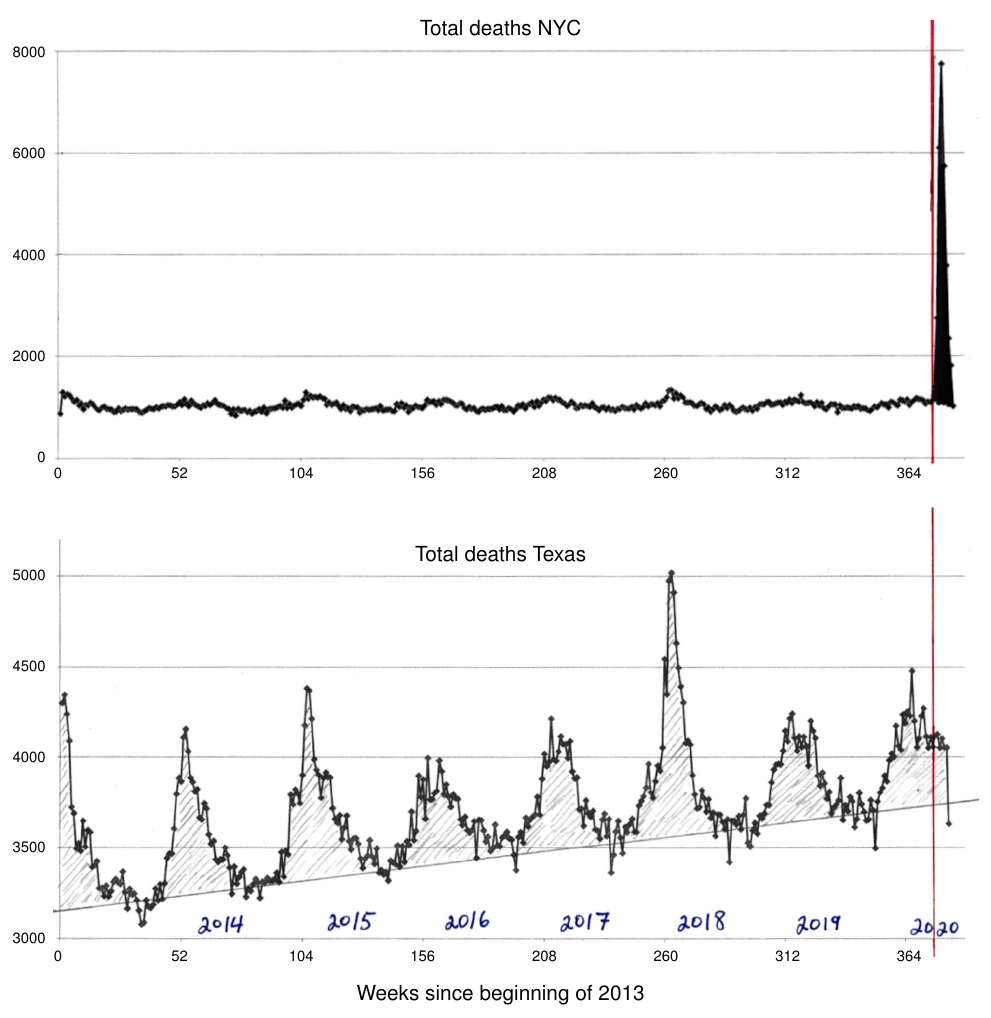
Figure 5: All-cause mortality in New York city and in Texas, before and after the declaration of the “pandemic” by the WHO (red vertical line). The x axis denotes the time in weeks since the beginning of 2013. A sharp yet short-lived spike occurred in New York City immediately after the declaration, whereas no such event was apparent in Texas. Note that, before the declaration, all-cause mortality for the winter 2019/20 had been tracking significantly lower than two years before; the higher mortality rate during that previous winter season was due to a worldwide outbreak of influenza that was not declared a pandemic. Graphics adapted from Figures 8 and 10 in Rancourt [23].

This, however, is demonstrably not the case: no relevant long-lasting increase in deaths from any cause (“all-cause mortality”) was observed for the winter of 2020. Rancourt has examined the correlation in time between the WHO’s declaration of the “pandemic” and all-cause mortality in various jurisdictions [23]. Immediately after the declaration, there occurred a sharp peak in all-cause mortality in some jurisdictions, but not in others; this discrepancy is illustrated in Figure 5 for New York City and Texas.

Common sense and historic precedents suggest that the spread of a deadly virus would not be stopped by international or state borders. Furthermore, if the virus had indeed been both deadly and truly novel, the wave of deaths in New York should not have subsided within such a short period of time as is apparent from Figure 5. The peak may instead signify that infected persons were transferred to New York City from other regions, were (mis)treated there, and died.

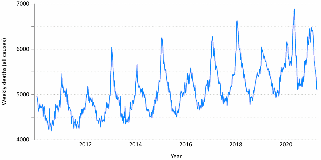
The mortality curve for the state of Texas shows no abnormalities for the winter of 2020, neither before nor after the WHO’s declaration of the pandemic. The same also applies to other jurisdictions such as Canada (Figure 6) and Europe. Thus, it is plain that in Winter 2020 there was no pattern of excess mortality that would reflect an usually severe and deadly virus pandemic.

no image info

Figure 6: All cause mortality in Canada by week, 2010 to 2021. Graphic from Rancourt et al. [24].

6. False assumptions serve as the basis for the calculations

The data used in the Watson et al. “modeling study” were based on hypotheses related to the Corona pandemic that have since been shown to be clearly wrong, namely:

  1. that there is no preexisting immunity and that the pandemic will affect everyone;
  2. that its effect can only be mitigated by political measures.

Ad 1: Serological investigations have shown that SARS-CoV-2 was endemic in Italy as early as September 2019 [25] and in France as early as November 2019 [26], which is thus likely true for Europe in general. However, at this time, no clustered disease activity was observed clinically. Antibodies to SARS-CoV-2 were found in up to 53% of asymptomatic individuals, whose serum samples had been collected in the pre-pandemic phase in Italy. In the meantime, more than 90 studies prove that a past infection with SARS-CoV-2 protects better against a recurrence of the disease than the vaccines [27]. While reinfections with new variants are possible, they are not severe [28]. These crucial facts are completely ignored by the authors. On the contrary, they speculate that possibly even more deaths than the estimated 14 million have been prevented by the “vaccination”, because in their calculations they may have underestimated the effect of the immune system’s failure to recognize new variants, which according to them would further increase the risk of a new infection.

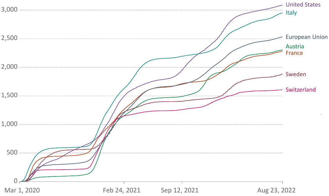
Ad 2: Over 400 studies show that non-pharmaceutical interventions such as lockdowns or school closures to prevent a pandemic are associated with no benefit, only harm [29]. Sweden, as one of the countries where hardly any restrictions were applied, performed significantly better in terms of the number of “COVID deaths” than many other countries with strict lockdown measures (Figure 7).

7. The claim of “vaccination success” is based on unscientific calculations

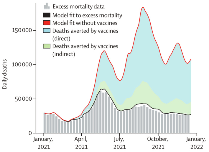
While Kyle Beattie’s estimate of hypothetical COVID mortality without vaccination is based on real data, namely on observations from countries with very low vaccination rates [11], Watson et al. use completely unrealistic and demonstrably false figures for their calculations to supposedly prove the efficacy of the COVID “vaccines.” This leads them to estimate an excessively high mortality which allegedly would have occurred without “vaccination.” From these inflated hypothetical death counts, they then subtract the officially reported deaths to obtain the “success of vaccination” (Figure 8). It is difficult to conceive of a more unscientific and misleading methodology than this one.

no image info

Figure 7: Cumulative confirmed COVID-19 deaths per million people, of various western countries as indicated. Graph retrieved from [30].
no image info

Figure 8: Global COVID-19 deaths prevented by vaccination, according to Watson et al. Mean number of daily “COVID-19 deaths” based on estimates of excess mortality (gray vertical bars) in the first year of vaccination. The baseline estimate of daily COVID-19 deaths resulting from fitting the model to excess mortality is shown as the solid black line, whereas the counterfactual scenario without vaccines is plotted as the red line. The distance between the red and black lines represents deaths prevented by vaccination. Among these, the proportion of deaths prevented by direct vaccine protection shown in blue and indirect protection in green. Adapted from Figure 1A in [1].

8. Serious conflicts of interest

If a publication raises as many questions as this one, then it is imperative to find out how it was financed. According to the declaration in the published paper itself, this “modeling study” was funded by, among others, the very institutions that earn millions from the “vaccinations”, thanks to an impressive return on investment ratio of 20:1 [31].

9. Conclusion

The hypothesis that the COVID “vaccinations” prevented 14 million “COVID deaths” and thus significantly mitigated the severity of the “pandemic” is based on unrealistic figures and demonstrably false calculations. To date, the experimental “vaccines”, for which an integration into the human genome cannot be safely ruled out [32,33] and must even be considered likely [34], have not been able to prove a relevant benefit, neither in the clinical studies before their approval nor afterwards. Real world evidence shows that the “COVID vaccination” is associated with a negative effect overall and positively correlates with an increase in morbidity and mortality associated with SARS-CoV-2 infection, as well as in all-cause mortality. Overall, the “modeling study” by Watson et al. must be considered an unscientific and dishonest attempt to falsely cast the “vaccines” in a positive light. The serious conflicts of interest surrounding the study undermine not only the credibility of the publication itself, but also that of the medical journal which published it.

Acknowledgement

We are grateful to Prof. Harald Walach, who made an important contribution to this opinion with his blog article [35].

References

  1. Watson, O.J. et al. (2022) Global impact of the first year of COVID-19 vaccination: a mathematical modelling study. Lancet Infect. Dis. (preprint)
  2. Polack, F.P. et al. (2020) Safety and Efficacy of the BNT162b2 mRNA Covid-19 Vaccine. N. Engl. J. Med. 383:2603-2615
  3. Baden, L.R. et al. (2021) Efficacy and Safety of the mRNA-1273 SARS-CoV-2 Vaccine. N. Engl. J. Med. 384:403-416
  4. Anonymous, (2020) A phase 1/2/3, placebo-controlled, randomized, observer-blind, dose-finding study to evaluate the safety, tolerability, immunogenicity, and efficacy of SARS-CoV-2 RNA vaccine candidates against Covid-19 in healthy individuals.
  5. Zaks, T. (2020) A Phase 3, Randomized, Stratified, Observer-Blind, Placebo-Controlled Study to Evaluate the Efficacy, Safety, and Immunogenicity of mRNA-1273 SARS-CoV-2 Vaccine in Adults Aged 18 Years and Older.
  6. Olliaro, P. et al. (2021) COVID-19 vaccine efficacy and effectiveness-the elephant (not) in the room. Lancet Microbe 2:e279-e280
  7. Alliance, C.C.C. (2022) Fact Checking The Fact Checkers.
  8. Latyopva, A. (2022) Did Pfizer Perform Adequate Safety Testing for its Covid-19 mRNA Vaccine in Preclinical Studies? Evidence of Scientific and Regulatory Fraud.
  9. Latypova, S. (2022) Moderna’s Non-clinical Summary for Spikevax—Evidence of Scientific and Regulatory Fraud.
  10. Data, O.W.i. (2022) Coronavirus (COVID-19) Deaths.
  11. Beattie, K.A. (2021) Worldwide Bayesian Causal Impact Analysis of Vaccine Administration on Deaths and Cases Associated with COVID-19: A Big Data Analysis of145 Countries. ResearchGate (preprint)
  12. Liu, Y. et al. (2022) Reduction in the infection fatality rate of Omicron variant compared with previous variants in South Africa. Int. J. Infect. Dis. 120:146-149
  13. Data, O.W.i. (2022) Coronavirus (COVID-19) Vaccinations.
  14. EuroMomo, (2022) Graphs and maps.
  15. Rancourt, D.G. et al. (2022) COVID-Period Mass Vaccination Campaign and Public Health Disaster in the USA. ResearchGate (preprint)
  16. Ioannidis, J.P.A. (2021) Reconciling estimates of global spread and infection fatality rates of COVID‐19: An overview of systematic evaluations. Eur. J. Clin. Invest. 5:e133554
  17. CDC, (2021) Coronavirus Disease 2019 (COVID-19) 2021 Case Definition.
  18. Corman, V.M. et al. (2020) Detection of 2019 novel coronavirus (2019-nCoV) by real-time RT-PCR. Euro Surveill. 25 (preprint)
  19. Zeichhardt, H. and Kammel, M. (2020) Kommentar zum Extra Ringversuch Gruppe 340 Virusgenom-Nachweis—SARS-CoV-2.
  20. Borger, P. et al. (2020) Corman-Drosten review report.
  21. Anonymous, (2022) Weekly Updates by Select Demographic and Geographic Characteristics.
  22. Anonymous, (2020) Report sulle caratteristiche dei pazienti deceduti positivi a COVID-19 in Italia. Il presente report è basato sui dati aggiornati al 17 Marzo 2020.
  23. Rancourt, D. (2020) All-cause mortality during COVID-19: No plague and a likely signature of mass homicide by government response. (preprint)
  24. Rancourt, D.G. et al. (2021) Analysis of all-cause mortality by week in Canada 2010-2021, by province, age and sex: There was no COVID-19 pandemic, and there is strong evidence of response-caused deaths in the most elderly and in young males.
  25. Apolone, G. et al. (2021) Unexpected detection of SARS-CoV-2 antibodies in the prepandemic period in Italy. Tumori 107:446-451
  26. Carrat, F. et al. (2021) Evidence of early circulation of SARS-CoV-2 in France: findings from the population-based “CONSTANCES” cohort. Eur. J. Epidemiol. 36:219-222
  27. Alexander, P. (2021) 91 Scientific Studies prove Naturally Acquired Immunity provides better protection than the Covid-19 Vaccines.
  28. Dhar, M.S. et al. (2021) Genomic characterization and epidemiology of an emerging SARS-CoV-2 variant in Delhi, India. Science 374:995-999
  29. Alexander, P. (2021) More Than 400 Studies on the Failure of Compulsory Covid Interventions (Lockdowns, Restrictions, Closures).
  30. Data, O.W.i. (2022) Cumulative confirmed COVID-19 deaths per million people.
  31. Belvedere, M.J. (2019) Bill Gates: My `best investment’ turned $10 billion into $200 billion worth of economic benefit.
  32. Zhang, L. et al. (2021) Reverse-transcribed SARS-CoV-2 RNA can integrate into the genome of cultured human cells and can be expressed in patient-derived tissues. Proc. Natl. Acad. Sci. U. S. A. 118 (preprint)
  33. Domazet-Lošo, T. (2021) mRNA vaccines: Why is the biology of retroposition ignored?. Preprint (preprint)
  34. Aldén, M. et al. (2022) Intracellular Reverse Transcription of Pfizer BioNTech COVID-19 mRNA Vaccine BNT162b2 In Vitro in Human Liver Cell Line. Curr. Issues Mol. Biol. 44:1115-1126
  35. Walach, H. (2022) Ohne Impfung 18 Millionen mehr Tote weltweit? – Wirklich? [Without vaccination 18 million more deaths worldwide? Really?].

Wie man ein Buch bespricht, ohne es zu lesen

Eine Antwort auf Peter Mayers Kritik an meinem Buch „Hiroshima revidiert“

Dr. Peter F. Mayer, Herausgeber der Online-Nachrichtenplattform tkp.at, schrieb kürzlich zwei kritische Artikel [1,2] zu meinem Buch Hiroshima revidiert [3].

Sollte er versucht haben, das Buch zu lesen, so war dieser Versuch wohl nicht von Erfolg gekrönt. Nach dem Inhalt seiner Artikel zu urteilen, gelangte er nicht über die dritte Seite hinaus. Und selbst diese sehr begrenzte Lektüre scheint ihm nicht gut bekommen zu sein. Das Ergebnis erinnert durchaus an Ephraim Kishons schöne Satire „Wie man ein Buch bespricht, ohne es zu lesen“. Wenn der Held von Kishons Geschichte dem Autor des betreffenden Buches über den Weg läuft und dieser die zugesagte Besprechung einfordert, zieht er sich aus der Affäre, indem er den Inhalt zu erraten versucht und sich dabei mehr und mehr in Rage redet. Allerdings gesteht er dem Autor am Ende, das Buch gar nicht gelesen zu haben. Auf eine solche Klarstellung hat Mayer bisher verzichtet.

Worin Mayer Recht hat

Mayers spontane und entschiedene Ablehnung meines Buches ist aber natürlich verständlich. Meine Kernthese – nämlich, dass in Hiroshima und Nagasaki keine Atombomben abgeworfen wurden, und dass die Amerikaner die speziellen Effekte von Atombomben lediglich vortäuschten – dürfte wohl den meisten Menschen abwegig erscheinen. Und wer sich dieser Überzeugung vergewissern möchte, dem steht dafür reichlich Material zur Verfügung. Für jeden Geschmack ist etwas dabei:

  • Berichte von Überlebenden, die den Atomblitz mit eigenen Augen sahen;
  • die Schatten angeblich verdampfter Körper von Menschen und Tieren auf dem Straßenpflaster;
  • viele Fälle von Strahlenkrankheit und Leukämie; und außerdem
  • viele seriös publizierte physikalische und medizinische Studien.

Mayer ist Physiker, und er konzentriert sich daher auf physikalische Studien, welche zeigen, dass die betreffenden Messproben starker Neutronen-Strahlung ausgesetzt gewesen sein müssen. Und wenn man diese Studien zum Nennwert akzeptiert, dann muss man tatsächlich zu der Schlussfolgerung gelangen, die Geschichte von den Atombomben in Hiroshima und Nagasaki sei korrekt – „case closed“.1

Widersprüchliche Beweise

Warum also schließe ich mich Mayers Sichtweise nicht an? Der Grund dafür ist „die andere“ Evidenz, welche der offiziellen Geschichte widerspricht. Ein Beispiel ist der Bericht des amerikanischen Militärarztes Paul Keller [4], den ich im ersten Kapitel meines Buches ausführlich zitiere. Keller berichtet von acht Überlebenden, die sich, nur von Holzhäusern abgeschirmt, nicht weiter als 500 Meter vom Zentrum der Detonation befunden hatten; oder genauer gesagt vom Hypozentrum, also dem Punkt am Boden, welcher senkrecht unterhalb der in der Luft gezündeten Bombe lag. Bei einem dieser Überlebenden soll diese Entfernung sogar nur 50 Meter betragen haben; er hätte sich also praktisch direkt unter der Bombe befunden.

Aber gemäß der offiziellen Geschichte hätten diese Menschen auf keinen Fall überleben dürfen! Und sie waren nicht die einzigen. Als man in späteren Jahren alle Überlebenden zu ihrem Aufenthaltsort während der Bombenangriffe befragte, um im Nachhinein ihre Strahlendosen zu schätzen, da tauchten noch mehr von ihnen auf [5]:

Obwohl einige Personen berichten, dass sie sich in geringer Entfernung [vom Hypozentrum] im Freien aufgehalten hatten, muss man ihre Geschichten als falsch ansehen, da die Intensität der Explosion und die Hitzeeinwirkung das Überleben in so geringer Entfernung unmöglich machten. … Dass sich so wenige Überlebende nicht an die Details des Ereignisses erinnern, lässt sich damit erklären, dass sie aufgrund der Explosion an Amnesie litten und ihre tatsächlichen Erfahrungen mit einer sie selbst zufriedenstellenden, erfundenen Geschichte ersetzt haben.

Mit anderen Worten: Überlebende, deren Erinnerungen nicht zur offiziellen Geschichte der Atombomben passen wollten, wurden von den Interviewern einfach als traumatisierte und verwirrte Spinner abgetan.

Wir haben also einerseits physikalische Messungen, die belegen, dass eine Atombombe losging; andererseits aber zeigt das Überleben dieser Betroffenen das Gegenteil. Es ist unmöglich, dass beide Beweise echt sind. Einer von ihnen muss gefälscht worden sein, und wir müssen zwischen ihnen wählen. Auf welcher Grundlage können wir das tun?

Welche Evidenz ist „hart“, welche ist „weich“?

Mayer macht es sich einfach, indem er dekretiert, dass „harte“ physikalischen Fakten die „weichen“ Fakten der Medizin grundsätzlich übertrumpfen. Aber diese Aussage ist ungerechtfertigt. In meinem Buch schrieb ich hierzu:

Alle physikalischen Dosimeter und Strahlungszähler sind Messfehlern unterworfen; aber kein ausgefranstes Kabel, keine lecke Batterie und keine Bedienungsfehler können die tödliche Wirkung von Strahlung auf den menschlichen Körper verhindern. … Wenn ein Mensch nicht stirbt, dann erhielt er keine tödliche Strahlendosis; es kann hierbei keine falsch-negativen Resultate geben. Wenn also eine physikalische Messung oder Berechnung ergibt, dass zu einer bestimmten Zeit und an einem bestimmten Ort tödliche Strahlung vorherrschte, aber andererseits ein Mensch überlebt, der genau dann und dort anwesend war, dann widerlegt dieses biologische Ergebnis kategorisch und endgültig die physikalische Aussage.

Es gibt noch eine zweite wichtige Überlegung: Welche der beiden einander widersprechenden Beweise ließe sich leichter fälschen?

Die Überlebenden ließen sich sicherlich nicht von interessierter Seite fälschen, und überdies hätte so etwas der offiziellen Geschichte nur unnötige Schwierigkeiten verursacht. Man kann auch kaum annehmen, dass die Betroffenen selbst ihre Geschichten eigens erfunden haben, um sich in den schon erwähnten Interviews als Spinner klassifizieren zu lassen. Es ist zudem interessant, dass Keller über die von ihm beobachteten acht Fälle ganz nüchtern und ohne zweifelnden oder abwertenden Kommentar berichtet. Vermutlich hatte man zu dem frühen Zeitpunkt seines Berichts (1946) noch keine klaren Sprachregelungen zur Frage des möglichen oder unmöglichen Überlebens verhängt, welchen er seine eigenen Eindrücke hätte unterordnen müssen.

Andererseits ist es technisch überhaupt kein Problem, Materialproben im Labor zu bestrahlen und sie dann ahnungslosen und wohlmeinenden Physikern als echte, von der Bombe bestrahlte Proben unterzujubeln.

Die hier angeführten Beispiele illustrieren das Prinzip, aber die verfügbare Evidenz erschöpft sich nicht in ihnen. Auch in anderen Fällen ergibt sich, dass die Beweise für die Bomben technisch leicht zu fälschen sind, die Beweise dagegen allerdings nicht.

Nobelpreisträger verwickeln sich in Widersprüche

Nun wird es sicherlich vielen Lesern widerstreben, den an der Entwicklung und Erforschung der Atombomben beteiligten Wissenschaftlern systematischen Betrug vorzuwerfen. In diesem Sinne äußert sich auch Mayer:

Physiker und Mathematiker wie Niels Bohr, Enrico Fermi, Hans Bethe, Eugene Wigner, John von Neumann, Richard Feynman, James Chadwick, Ernest Lawrence, Isidor Isaac Rabi, Wolfgang Pauli und einige weitere Nobelpreisträger Lügner und Betrüger zu nennen, ist ein starkes Stück.

Ich kann Mayer an diesem Punkt durchaus verstehen – auch mir selbst ging dies anfangs gegen den Strich. Aber wiederum sprechen die Fakten für sich selbst. Bevor wir uns diesen zuwenden, muss ich noch anmerken, dass Mayers umfangreiche Namensliste von ihm selbst zusammengestellt wurde – sie findet sich nicht in meinem Buch, welches er ja auch gar nicht kennt.

Nun zu den Fakten. Einige davon finden sich im Protokoll des Treffens einer geheimen Kommission [6]. Das Treffen dieses sogenannten „Interim-Komitees“ fand am 31. Mai 1945 in Washington, DC statt. Neben Politikern, Offizieren, und einigen anderen schillernden Figuren waren auch die folgenden Wissenschaftler anwesend: Arthur und Karl Compton, James Conant, Robert Oppenheimer, Enrico Fermi und Ernest Lawrence. Ganz oben auf der Agenda war der gegenwärtige Entwicklungsstand der Atombomben. Arthur Compton fasste diesen wie folgt zusammen:

  1. Die Anreicherung von Uran-235 wird bereits beherrscht, und Uran-Bomben befinden sich „in Produktion“.
  2. Plutonium-Bomben sind erstrebenswert, und man erwartet von ihnen eine erheblich größere Sprengkraft als von Uran-Bomben.
  3. Die Produktion von Plutonium steht aber noch ganz am Anfang. Bisher ist es lediglich gelungen, durch den Betrieb von Uran-Reaktoren das Rohmaterial hierfür zu erzeugen, d.h. eine komplexe Mischung von radioaktiven Isotopen, welche auch Plutonium enthält. Die Reinigung von Plutonium aus dieser Mischung muss erst noch entwickelt werden.
  4. Diese Entwicklung wird noch etwa zwei Jahre erfordern. Danach werden noch weitere 18 Monate vergehen, bis Plutonium „in Menge“ erhalten werden kann.

Enrico Fermi teilt mit, dass es ihm derzeit an ausreichend Rohmaterial fehlt, um die Entwicklung der Plutonium-Reinigung voranzutreiben. Oppenheimer spricht davon, dass eine Uran-Bombe getestet werden soll, um deren Explosionsenergie zu messen; er erwartet ein Resultat im Bereich von 2000 bis 20.000 Tonnen TNT.

Keiner der anwesenden Wissenschaftler gibt irgendwelche Vorbehalte gegen die Aussagen Comptons, Fermis oder Oppenheimers zu Protokoll. Aber diese Aussagen widersprechen der allgemein bekannten Atombomben-Legende in mehreren zentralen Punkten. Gemäß dieser Legende

  1. fand der von Oppenheimer dem Interim-Komitee angekündigte Test einer Uran-Bombe nie statt. Ein solcher Test war angeblich nicht notwendig;
  2. wurde nur eine einzige Uran-Bombe gezündet, nämlich die in Hiroshima, obwohl solche Bomben (Mehrzahl) ja angeblich „in Produktion“ waren;
  3. wurden in Nagasaki sowie bei dem Test in Alamogordo zwei Plutonium-Bomben eingesetzt, obwohl ja laut dem Protokoll des Interim-Komitees Plutonium noch gar nicht verfügbar war.

Mir ist nicht bekannt, dass irgendeiner der hier genannten Wissenschaftler jemals dieser offiziellen Legende öffentlich widersprochen hätte. Schon anhand dieses einen offiziellen Dokuments können wir also feststellen, dass diese Wissenschaftler die Täuschung der Öffentlichkeit zumindest schweigend hingenommen haben. Mehrere von ihnen wirkten auch nach dem Krieg weiterhin aktiv daran mit; siehe zum Beispiel das Propaganda-Pamphlet “One World Or None” [7].

Die Einsicht, dass diese Geistesriesen in ethischer Hinsicht eher Zwerge waren, mag Mayer ähnlich hart treffen wie seinerzeit die Entzauberung des Weihnachtsmanns, sie ist aber gleichermaßen unvermeidlich.

Fazit

Ich habe hier bewusst darauf verzichtet, im Detail auf die vielfältige Evidenz einzugehen, die in meinem Buch zu finden ist. Stattdessen habe ich nur versucht zu zeigen, dass

  • die in meinem Buch gestellten Fragen berechtigt sind,
  • sich diese Fragen nicht anhand einseitig ausgewählter Evidenz beantworten lassen,
  • die mit diesen Fragen befassten wissenschaftlichen und politischen Autoritäten unser blindes Vertrauen nicht verdienen.

Ebenso wie Richter und Geschworene die einander widersprechenden Beweismittel beider Parteien kritisch bewerten müssen, so müssen auch wir bei den uns überlieferten Daten und Legenden zu Hiroshima und Nagasaki sorgfältig die Spreu vom Weizen trennen.

Genau dies habe ich mit meinem Buch versucht. Dabei hatte ich die Unterstützung von Spezialisten anderer Disziplinen. So wurde zum Beispiel das Kapitel zum Fallout von einem Physik-Professor kritisch gelesen und verbessert, der selbst mehrere Bücher über die Funktionsweise und die Geschichte von Atombomben verfasst hat. Ein Augenarzt und ein japanischer Spezialist für Hautverbrennungen (Dr. Teruichi Harada) überprüften die medizinischen Aussagen zu ihren Fachgebieten.

Wie eingangs erwähnt, komme ich zu dem Schluss, dass in Japan keine Atombomben detoniert worden sein können. Dieses Ergebnis erfordert letztlich auch eine neue Deutung der Motive Amerikas. Auch dies habe ich versucht. Weder hierbei noch bei den wissenschaftlichen Fragen erhebe ich Anspruch auf Unfehlbarkeit. Es würde mich aber durchaus freuen, wenn zukünftige Kritiker zunächst meine Argumente zur Kenntnis nähmen, bevor sie sie verwerfen.

1 Zusätzlich bringt Mayer auch noch einige sehr viel weniger zwingende physikalische Argumente vor, aber es ist unnötig, die Diskussion über diese hier auszubreiten.

Literatur

  1. Mayer, P.F. (2025) Warfen die USA 1945 Atombomben auf Hiroshima und Nagasaki ab?. URL: https://tkp.at/2025/08/12/warfen-die-usa-1945-atombomben-auf-hiroshima-und-nagasaki-ab
  2. Mayer, P.F. (2025) Atombomben auf Hiroshima und Nagasaki – Fakten und Fiktionen. URL: https://tkp.at/2025/08/18/atombomben-auf-hiroshima-und-nagasaki-fakten-und-fiktionen/
  3. Palmer, M. (2024) Hiroshima revidiert (Etica Media). URL: https://eticamedia.eu/produkt/hiroshima/
  4. Keller, P.D. (1946) A clinical syndrome following exposure to atomic bomb explosions. J Am Med Assoc 131:504-6. URL: https://www.ncbi.nlm.nih.gov/pubmed/?term=20983706
  5. Jablon, S. (1971) Atomic bomb radiation estimation at {ABCC} (Atomic Bomb Casualty Commission). URL: https://www.rerf.or.jp/library/scidata/tr_all/TR1971-23.pdf
  6. Arneson, R.G. (1945) Notes of the Interim Committee meeting, Thursday, 31 May 1945. URL: https://archive.org/details/interim-committee-may31-1945
  7. Masters, D. und Way, K. (1946) One World Or None: A Report to the Public on the Full Meaning of the Atomic Bomb (New Press). URL: https://archive.org/details/oneworldornonere00mast

中性子放射線に関する和中光次氏の投稿への反論

日本のブロガーである和中氏は、彼の最近の投稿の中で、広島と長崎では原子爆弾が爆発しなかったと論じている私の著書『Hiroshima Revisited』を批判している。和中氏は特に第6章に焦点を当てており、広島の原爆によって放出されたとされる中性子放射線に関する物理学的研究について論じている。その論理や根拠は以下の通りである:

  1. 爆弾から放出され地面に到達した中性子は、そこにある原子核にランダムに吸収される。
  2. 中性子を吸収した原子核の一部は、それによって放射性となる。
  3. 後日、そうした原子核の放射能を測定することで、爆発によってどれほどの中性子放射線が放出されたかを特定することができる。

問題となる放射性原子核は、それぞれ異なる元素や同位体に属しており、それぞれに特有の半減期やその他の性質を持っている。それらの同位体の相対的な存在比率から、原爆中性子のエネルギースペクトルに関して、いくつかの結論を導き出すことができる。さらに、爆発地点からの距離が異なる場所で採取された試料を比較することで、原爆中性子がどれだけの距離を移動したかを特定することができる。

以上が理論的な説明である。さて、こうした研究(つまりDS02)の「研究前提そのもの」が、明らかに私の著書の中心的な主張と相容れない。もし実際に原子爆弾が爆発していなかったとしたら、中性子によって誘発された放射線は一体どこから来たのだろうか? 私は、中性子照射を受けた試料はすべて、まさにこうした研究のために捏造されたものだと主張している。そしてここで、極めて重要な点を強調しておかなければならない:

私が中性子研究を偽造であると断定したのは、その研究の内部にある矛盾のためではなく、研究の範囲とはまったく無関係な証拠に基づいてのことである[1]。

その証拠の一つとして、爆心地付近で、本来なら生存不可能とされるレベルの放射線を浴びたにもかかわらず、生き残った人々が相当数存在することが挙げられる。私の著書にも関連する統計データが掲載されているが、林千勝氏の『広島・長崎 悲劇の正体』では、より説得力のある詳細情報や当事者の証言が掲載されている。さらなる理由や中性子放射線に関する研究などよりも、「そこで生存できたという事実」を優先すべき理由については、私の著書またはその日本語訳『偽装された原爆投下』を参照されたい(こうしたケースでの科学的重みづけとしては、直接的効果が優先され、後日の測定などの間接的効果の評価エビデンスレベルは落ちる)。

和中氏は(DS02と同様に)この前提を無視しているため、自身の分析全体を、誤った前提から始めてしまっている。彼は次のように書いている。

1990年代には、「DS86中性子問題」が議論された。広島の爆心地から遠く離れた場所のサンプルから、理論からの予想計算値を上回る放射線量が測定されたというものである。これは無視しえない学術的争点である。パルマーはこの不一致を、データが捏造されたという主張の出発点として利用している[2]。

和中氏は、私の著書の該当するくだりを、私が重視している文脈から切り離して解釈し、「その目的が中性子データの捏造を証明することにある」と想定している。しかし、私の真意は、単にそれらがどれほど巧妙に捏造されたかを検証するだけのことである。したがって本章全体の意義とは、本書の中心的な論旨(被害の直接効果が原爆出力に合わないこと)に対する単なる脚注に過ぎない。

ここで中性子研究の内容に焦点を当てると、私自身の主張と和中氏の主張の違いは、次のように要約できる。

  • 私は、中性子放射線に関する古くからの一連の研究には、解決できない矛盾が数多く含まれていると主張している。
  • 和中氏は、古い時代の矛盾は重要ではないと主張している。中性子放射線に関する最新の研究のみを有効とみなすべきであり、それ以前のものはすべて無視すべきだという。

繰り返すが、ここで問題となっているのは、捏造手段の巧妙さだけである。私は、それが露見していると主張する。もし和中氏が、中性子線が存在したと上手く述べられたとしても、それは研究が「より巧妙に捏造されていた」ことを意味するだろう。実際のところ、彼はDS02の主張を証明していない。これは後述部分で明らかになるだろう。

中性子放射線に関する研究における中心的なパラメータの一つが緩和長(減衰長)であり、これは中性子がその経路上の原子核に「吸収」されるまでに、平均して空気中を、どれだけの距離を移動できるかを表している。1950年代にネバダ州で開始された実験研究により、緩和長は235メートルと算出されていた。広島と長崎の異なる大気条件下では、198メートルという値が想定された。その後、両都市から採取された試料における中性子誘発放射能の測定により、この値が裏付けられた。それにもかかわらず、1980年代初頭、「緩和長を
155メートルに短縮すべき」とする新たな理論モデルDS86が提唱された。当然のことながら、この新しい理論による推定値は、既存の測定結果と矛盾していた。さらに、その提唱後に実施された測定研究でさえ、新しい理論ではなく、従来の理論を引き続き支持する結果となった(もちろんサンプルは当時の古いもの)。

このような状況下で、まともな科学者ならどうするだろう? 研究結果が合っていないことを認め、自らの理論がエビデンスによって否定されたことを受け入れ、より優れた改良された理論を探すはずだ。しかしながらこのケースでは、わざわざ探す必要すらなかった。単に古い理論に戻ればよかっただけなのだ!

しかし、広島のデータを担当する科学機関はそうしなかった。その代わりに、提唱後から
10年以上にわたって適合する証拠が一切得られないままに、頑なに新しい理論に固執し続けた。つまり彼らが追求していたのは科学的真実ではなく、政治的な意図に合わせる行為だったのは明らかだ[3].。

安堵が訪れたのは2000年代初頭になってからで、理論計算と実験データの両方に合うように、「新しく改良された」バージョンに更新された公式報告書が発表された時だった。これが、和中氏が自身の投稿で取り上げているDS02報告書である[4]。この報告書における新たな計算は、「大した内容ではない」と評することができる。また以前の理論からの変更点はごくわずかであり、両バージョンを区別するために利用可能な実験データの精度も極めて低い。そのため、なぜこれらの新たな計算を作成し、公表する価値があると考えられたのか不思議に思える。私の推察では、それらが「新しく改良された」証拠を公表するための、単なる口実に過ぎなかったのではないかと疑っている。

それでは、その証拠について見てみよう。和中氏の投稿では、中性子放射線によって生成される3つの放射性同位体(塩素36、ユーロピウム152、コバルト60)に関する測定結果が取り上げられている。いずれの場合も、彼は、旧説と一致していた従来の測定値は破棄し、新説に合致する新しい測定値を受け入れるべきだと主張している。基本的な論点はどのケースでも同じである。すなわち以前の研究では、測定信号に含まれるある種の自然背景放射線の寄与(上乗せ)が見落とされており、それが誤って爆弾の放射線として計上されていたという。爆発地点から離れるほど、こうした背景放射線の割合は大きくなるため、爆発地点に近い場所で採取された試料と比較して、測定値は遠位で過大に表示されてしまう。その結果、緩和長、すなわち中性子の到達距離の推定値が過大評価されてしまうことになる。

和中氏の主張に妥当性があるかどうかを判断するには、次の点を問わなければならない。すなわち、先行研究の著者たちは、バックグラウンド放射線を「本当に考慮に入れてなかったのか?」、それとも「本当は考慮していたのか?」。詳細な議論に入る前に、2つの一般的な指摘をしておきたい:

  1. 研究者たち(しかも単一の研究者ではなく、数十年にわたる研究活動を通じて複数の研究グループ)が、そのような重大な過ちを犯し続けていたという発想そのものが荒唐無稽である。実験研究のための教育を受け実際に施行した経験を持つ者なら、誰一人として、そんなお粗末な失敗があったなどと考えもしないだろう。
  2. それらの研究がバックグラウンド放射線を考慮していたかどうかを検証するには、当然ながら論文を全文読む必要がある。しかし、和中氏は次のように述べている。「1992、2005年、2008年の論文については、要旨のみを検討した。論文の全文は検討していない」。 これは、彼が自身のブログ記事で引用している論文に関する記載である。おそらく彼は、私の著書で引用されている研究論文も一切読んでいないのだろう。

初期の論文を否定する前に、もしもいくつかの論文を実際に調べてみれば、以前の研究者たちが背景放射線の問題を十分に認識していたことがすぐにわかるだろう。コバルト
60放射線に関する最初の論文は、1967年に橋爪らによって発表された。そこには次のように記されている[5]:

したがって、分光器のバックグラウンド(自然放射線などの、いわゆるノイズ)は、
GM管のバックグラウンドによって制限され、宇宙線粒子(ミューオン)によるバックグラウンドとの識別は、パルス高の選別によって行われる。この種の検出器の詳細な性能については、すでに報告されている16-18。

その後、コバルト60放射線を調査したすべての研究者は、この初期の研究に精通していたと推測できる。そして、さらに改善しようと試みる中で、同様にバックグラウンド放射線を考慮し、補正するために最善を尽くしたはずである。これらの初期の研究者たちは、
DS02報告書が作成された当時よりも精巧さの落ちた測定機器を用いざるを得なかったものの、信号対雑音比が高いという利点には恵まれていた。例えば、1967年から
2000年の間に、核爆発によって生成されたコバルト60の放射能は、およそ75分の
1まで減衰していたはずである(つまりノイズに対してコバルト60の放射能は高かったので、主信号の検出は後の時代よりも容易だったはずだ)。

塩素36の初期の測定結果は、1992年にストラウメらによって報告された[6]。和中氏は、これらの著者らが考慮していなかったとされる塩素36の自然バックグラウンドを大いに問題視している。しかし、この研究の「表1」には、正確に1708メートルの「傾斜距離」で採取された4つの試料が記載されている。これは地上距離で爆心地から1606メートルに相当し、調査対象となった全試料の中で最も遠い距離である。したがって、「バックグラウンド放射線への無視が誤った測定値を生んだ」と指摘される点に関して、最も重要な資料になる。4つの試料はすべて同じ建物から採取されたもので、そのうち2つの試料は、爆弾の放射線に被曝したと記述されている。これらの試料について、安定同位体である塩素35に対する塩素36の存在比は、それぞれ579 × 10-15および310 × 10-15と示されている。残りの2つの試料は「完全に遮蔽されていた」と記述されており、表の中では明確に「バックグラウンド」に該当する資料だと記載されている。測定値は、それぞれ120 × 10-15および
125 × 10-15である。明らかに、ストラウメとその共著者たちもバックグラウンドの問題を認識しており、それに対処していたのである。

ユーロピウム-152に関する研究において、バックグラウンド放射線を無視したとして、後に非難された研究者を引用させてほしい。それは静間清であり、彼は3人の同僚と共に、まさにバックグラウンド放射線を最小化するための研究を発表していた[7]。 この研究の要旨を次のように述べている:

原爆中性子によって誘起された残留Eu152の放射能を測定するため、低バックグラウンドのガンマ線分光計が作製された。通常の同軸型およびウェル型のGe検出器について、鉛遮蔽材の最適厚さ、内張り、およびバックグラウンド特性を調査した。さらに、ウェル型検出器にはアンチコインシデンス遮蔽を設置した。その結果、宇宙線に起因するバックグラウンド計数率は大幅に低減された。また、Eu152の測定においては、対象となる放射能物質を濃縮して、バックグラウンド放射能を除去するための試料調製が重要であることが分かった。

私はこの技術的操作の妥当性を明らかにするために、この分野を専門とするドイツ人研究者に連絡して相談した。「静間氏は一貫して、極めて慎重かつ正確に研究を行った」と賞賛している。これはまさに、静間氏の論文を読んだ際に受ける印象そのものである。これまでの話を要約すると、「静間氏やその他の先行研究者たちがバックグラウンド放射線を無視していた」とするDS02報告書の主張は、荒唐無稽なだけでなく中傷的ですらある。和中氏は自説としてDS02の主張を肯定する前に、こうした内容を吟味すべきだった。DS02と同じ主張を繰り返すべきではなかった。

和中氏はユーロピウム152に関して、そのガンマ線のエネルギーがアクチニウム
227のガンマ線と類似しているため、区別できないとも主張しているが

  1. これら2つの同位体のシグネチャ(その同位体に特徴的な信号など)を区別できる検出器は、かねてより利用可能であった。
  2. アクチニウム227は半減期の短い同位体であり、ウラン235およびトリウム232の崩壊系列の一部としてのみ存在するため、常にそれらの系列に含まれる他の同位体と共に検出される。したがって、たとえその放射能がユーロピウム152の放射能によって覆い隠されたとしても、他の同位体のシグネチャに基づいて計算し、補正することが可能である。

もう一つの選択肢は、同位体組成を測定する前に、試料からユーロピウムを化学的に抽出することである。実際、静間らによる研究(上記の引用参照)では、この方法が採用された。したがって、和中氏(DS02)が指摘した問題点は対処可能であり、実際に解決されてもいた。

以上の点は、後に論争の的にされたバックグランド問題が、すでに解決されていたことを証明している。古い研究では実際にバックグラウンド放射線が考慮されていたため、DS02報告書で古い時代の測定値を無効にした理由は破綻している。したがって、古い実験データセットと新しい実験データセットの間には、未解決の矛盾が残されていることになる。私の著書の第6章では、和中氏が論じた点よりも、さらに顕著な矛盾をいくつか挙げている。科学的に厳密に言えば、これらの矛盾は、データの「一部」が捏造されたことを証明するに過ぎず、「すべて」が捏造されたとまでは証明できない。冒頭で指摘したように、後者の主張は、こうした研究が言及できる範疇を超えた領域の議論になってしまう。

和中氏は、私の記述内にその他にも誤りがあるとして挙げている。しかし、それらは上述事項と同様に、彼自身の誤謬や誤解に起因するものである。それらについて詳細に論じることには、読者の時間を割く価値がないと考える。したがって和中氏に対しては、次回はまず下調べをしっかりと行うよう勧めて、この文章を締めくくりたい。

注釈および参考文献

[1] 少々陳腐な例えではあるが、ある警察官が、X氏がY氏を「同日に銀行強盗を犯した」と告発しているのを聞いている場面を想像してほしい。その警察官は、前日に別の銀行強盗の容疑でY氏を逮捕していたため、X氏の証言の詳細を検討しなくても、X氏が嘘をついていることは分かる。

[2] 和中氏の投稿からのすべての直訳引用は、DeepLを使用して翻訳した。

[3] この一連の出来事の背景にあったであろう動機については、私の著書の第11章で扱っている。

[4] Young, R.W. and Kerr, G.D. (2002) Reassessment of the atomic bomb radiation dosimetry for Hiroshima and Nagasaki: dosimetry system 2002 https://www.rerf.or.jp/library/scidata/scids/ds02

[5] Hashizume, T. et al. (1967) Estimation of the air dose from the atomic bombs in Hiroshima and Nagasaki. Health Phys. 13:149-61 https://www.ncbi.nlm.nih.gov/pubmed/?term=6029426

[6] Straume, T. et al. (1992) Neutron discrepancies in the {DS}86 Hiroshima dosimetry system. Health Phys. 63:421-6 https://www.ncbi.nlm.nih.gov/pubmed/?term=1526783

[7] Shizuma, K. et al. (1992) Low-background shielding of Ge detectors for the measurement of residual 152Eu radioactivity induced by neutrons from the Hiroshima atomic bomb. Nucl. Instrum. Methods Phys. Res. B 66:459 – 464
http://www.sciencedirect.com/science/article/pii/0168583X9295419R

The truth about the bombings of Hiroshima and Nagasaki

This page presents some of the evidence contained in my book Hiroshima revisited. It makes the case that the bombings of Hiroshima and Nagasaki were not atomic. It presents two kinds of evidence—namely, physical and medical—which both contradict the story of the atomic bombings. The medical evidence additionally indicates that “radiation sickness” was faked with mustard gas, whose biological effects resemble radiation, and flash burns were faked with napalm. The political and historical context of the fake atomic bombings is briefly considered here as well.

I have also covered about the same ground in a video. My friend Dr. Teruichi Harada has recorded a Japanese version of that video.
Continue reading

Rebuttal of Mr. Mitsui Wanaka’s post regarding “Hiroshima Revisited” and neutron radiation

In a recent post, Japanese blog writer Mitsui Wanaka criticizes my book Hiroshima Revisited, which makes the case that no nuclear bombs were detonated in Hiroshima and Nagasaki. Mr. Wanaka focuses in particular on the sixth chapter, in which I discuss physical studies on the neutron radiation that was supposedly released by the Hiroshima bomb. The rationale of such studies is as follows:

  1. Neutrons from the bomb that strike the ground will be taken up at random by atomic nuclei there.
  2. Some of the nuclei that took up a neutron will thereby become radioactive.
  3. We can measure, at some later time, the radioactivity of such nuclei in order to determine how much neutron radiation was released by the detonation.

The radioactive nuclei in question will belong to different chemical elements and isotopes, each with their own characteristic half-lives and other properties. From the relative abundance of those isotopes, we can draw some conclusions regarding the energy spectrum of the bomb neutrons. Furthermore, by comparing samples collected at different distances from the detonation, we can determine how far the bomb neutrons had traveled.

Thus far the theory. Now, the entire premise of such studies is obviously incompatible with the central thesis of my book. If indeed no atomic bombs were detonated, then where does the neutron-induced radiation come from? I posit that all of the neutron-irradiated samples were forged for the sake of these very studies. And here I must emphasize a crucial point:

I ruled the neutron studies to be fake not because of their internal contradictions, but based on evidence entirely out of their own scope.1

A straightforward piece of such evidence is the considerable number of people who survived supposedly unsurvivable radiation near the hypocenter. My own book contains some relevant statistics, but Chikatsu Hayashi’s more recent book (in Japanese) has more compelling detail and first-hand testimony. For further evidence, and for the reasons to prefer it to studies on neutron radiation etc., please see my own book or its Japanese translation.

Because Mr. Wanaka ignores this background, he starts his entire analysis from a mistaken premise. He writes:

In the 1990s, the “DS86 neutron problem” was discussed, in which samples far from the epicenter in Hiroshima had radiation measurements that exceeded calculations. This is a real academic debate. Palmer uses this discrepancy as a starting point for his claim that the data was fabricated.2

Mr. Wanaka, taking my book chapter out of context, assumes that its purpose is to prove that the neutron data were faked. However, its real purpose is merely to examine how skilfully they were faked. The entire chapter thus is merely a footnote to the central thesis of the book.

Focusing now on the substance of the neutron studies, the difference between my own arguments and Mr. Wanaka’s can be summed up as follows:

  • I argue that the collective studies on neutron radiation contain numerous contradictions that cannot be resolved.
  • Mr. Wanaka argues that these contradictions do not matter—only the most recent studies on neutron radiation should be considered valid, and all previous ones should be ignored.

Again: the only thing at stake here is the cleverness of the fraud. I posit that it is transparent. If Mr. Wanaka could prove his point, this would mean that the studies had been faked rather more proficiently. However, he has not proved his point. This will become clear below.

One central parameter in the studies on neutron radiation is the relaxation length, which determines how far, on average, the neutrons can travel through the air before being “swallowed up” by some atomic nuclei in their path. Experimental studies begun in Nevada during the 1950s had yielded a relaxation length of 235 meters. Under the different atmospheric conditions in Hiroshima and Nagasaki, a value of 198 meters was assumed. Subsequent measurements on neutron-induced radioactivity in samples from both cities confirmed this value. Nevertheless, in the early 1980s, a new theoretic model was introduced, according to which the relaxation length should be reduced to 155 meters. The new theory thus contradicted the available measurements. What is more, the measurements conducted after its introduction continued to support the old theory but not the new one.

What would a real scientist do in such a situation? He would admit defeat—accept that his theory is rejected by the evidence and look for a better one. And in this case, he would not even have to look at all, since he could simply go back to the old theory!

But this is not what the scientific institutions in charge of the Hiroshima data did. Instead, they stubbornly clung to the new theory, even though for more than a decade no supporting evidence was forthcoming. They obviously were not pursuing scientific truth but rather a political agenda.3

Relief came only in the early 2000s with an updated official report that contained “new and improved” versions of both the theoretical calculations and the experimental data. This was the DS02 report,4 which Mr. Wanaka highlights in his post. The new calculations in this report can be described as a “nothing-burger.” The changes from the previous theory are so minor, and the accuracy of the experimental data available to distinguish between the two versions is so low, that one wonders why anyone found it worthwhile to prepare and publish these new calculations. I suspect that they were only a pretext for unveiling the “new and improved” evidence.

Let us now take a look at the evidence. In his post, Mr. Wanaka deals with measurements on three radioactive isotopes induced by neutron radiation: chlorine-36, europium-152, and cobalt-60. In each case, he argues that we should discard the old measurements, which agreed with the old theory, and accept the new ones, which fit the new theory. The basic argument is the same in each case—namely, that the old studies had overlooked the contribution of some kind of natural background radiation to the measured signals, which they had mistakenly included in the bomb radiation. Because the proportion of such background radiation will be greater at larger distances from the detonation, these will appear too high relative to samples obtained nearer the detonation. The result will be an inflated estimate of the relaxation length, i.e. of the neutrons’ reach.

To decide if Mr. Wanaka’s claim has merit, we must ask: did the authors of those earlier studies fail to account for background radiation, or did they not? Before we go into details, two general remarks are in order:

  1. The whole idea that researchers—and not just single researchers, but multiple groups of them, throughout decades of research work—would commit such a blunder is absurd. Nobody with any training and experience in experimental research would believe it for a minute.
  2. To verify whether or not those studies accounted for background radiation, one would obviously have to read them in their entirety. However, Mr. Wanaka notes: “For the 1992, 2005, and 2008 papers, the abstracts were reviewed. The full texts of the papers were not reviewed.” This pertains to the papers he cites in his own blog post. I assume that he did not read any of the research papers cited in my book either.

If we do trouble to actually examine some of the early papers before dismissing them, we quickly see that the authors were fully aware of the background radiation problem. The very first paper on cobalt-60 radiation was published by Hashizume et al. in 1967. There, we read:5

The background of the spectrometer is thus restricted by that of the GM tube, and discrimination against a cosmic ray particle background (muons) is made by pulse height selection. The detailed performance of a detector of this type has already been reported.16-18

We can assume that all later investigators who examined cobalt-60 radiation were familiar with this early study and, trying to improve on it, would likewise have done their best to account and correct for background radiation. While these early investigators had to make do with less sophisticated instruments than were available at the time of the DS02 report, they did have the benefit of a higher signal-to-noise ratio. For example, between the years 1967 and 2000, the activity of bomb-induced cobalt-60 would have decayed by a factor of about 75.

Early measurements of chlorine-36 were reported by Straume et al. in 1992.6 Mr. Wanaka makes much of the natural background of chlorine-36 that was allegedly not accounted for by these authors. However, Table 1 of Straume’s study lists four samples that were obtained at a “slant range” of exactly 1708 meters. This corresponds to a ground distance of 1606 meters—the largest distance of all samples examined, and therefore the most relevant to the alleged neglect of background radiation. All four samples came from the same building. Two samples are described as exposed to the bomb radiation. With these samples, the measured abundance of chlorine-36, relative to the stable isotope chlorine-35, is given as 579×10-15 and 310×10-15, respectively. The two other samples are described as “totally shielded”, and the table explicitly lists them as “background.” The measured values are 120×10-15 and 125×10-15, respectively. Evidently, Straume and his co-authors, too, were familiar with the background problem and dealt with it.

Regarding the studies on europium-152, let me cite one of the authors that were accused later on of having neglected background radiation. Kiyoshi Shizuma, together with three colleagues, published a study whose very purpose was the minimization of background radiation.7 The abstract of this study reads:

Low-background gamma-ray spectrometers were constructed for the measurement of residual 152Eu activity induced by the atomic-bomb neutrons. Optimum thickness of lead shielding, inner linings and background characteristics were investigated for an ordinary coaxial- and a well-type Ge detector. In addition, an anticoincidence shielding was installed for the well-type detector. As a result, the background counting rate due to cosmic rays was greatly reduced. It was also shown that a sample preparation to enrich the objective activity and eliminate background activities was important in the case of the 152Eu measurement.

A German researcher in this field whom I had contacted to clarify some technical points volunteered that “Shizuma’s work was always extremely careful and accurate.” This is precisely the impression one gets when reading Shizuma’s papers. In summary, the allegations that Shizuma and all those other earlier researchers had neglected background radiation are nonsensical and scurrilous. Mr. Wanaka should have known better than to repeat such allegations without vetting them first.

Mr. Wanaka also claims that the gamma radiation of europium-152 cannot be distinguished from that of actinium-227, which is similar in energy. However,

  1. detectors that can discriminate the two isotopes’ signatures have been available for a long time,
  2. actinium-227 is a short-lived isotope that occurs only as part of the uranium-235 and the thorium-232 decay chains, and thus is always accompanied by the other members of those chains. Therefore, even if its own activity were obscured by that of europium-152, it could be calculated and corrected for based on the signatures of those other isotopes.

Another option is to chemically extract europium from the sample before determining its isotopic composition. This was indeed done in the study by Shizuma et al. (see quote above). The difficulty raised by Mr. Wanaka therefore can and has been dealt with.

The above points address the major bone of contention. Since the older studies did in fact account for background radiation, the reason given by Mr. Wanaka for dismissing them is invalid. We are thus left with unresolved contradictions between the older and the newer experimental data sets. My book chapter cites several other, even more glaring contradictions than those discussed by Mr. Wanaka. Strictly speaking, these contradictions prove only that some of the data were faked; they cannot prove that all of them were. As pointed out above, the latter assertion rests on evidence outside the scope of these measurements themselves.

Mr. Wanaka alleges several other supposed errors on my part which, just like the ones discussed above, result from errors and misunderstandings of his own. I do not consider it worth the time of my readers to address them in detail. I will therefore close with the recommendation to Mr. Wanaka to first do his homework next time.

Notes

  1. For a somewhat trite analogy, consider a policeman listening to Mr. X accusing Mr. Y of robbing a bank on the same day. Since the same policeman had arrested Mr. Y on the day before for robbing another bank, he does not need to consider the details of Mr. X’s testimony to know that Mr. X is lying. (back)
  2. All literal quotes from Mr. Wanaka’s post were translated using DeepL. (back)
  3. I deal with the likely motive for this maneuver in Chapter 11 of my book. (back)
  4. Young, R.W. and Kerr, G.D. (2002) Reassessment of the atomic bomb radiation dosimetry for Hiroshima and Nagasaki: dosimetry system 2002 https://www.rerf.or.jp/library/scidata/scids/ds02/ (back)
  5. Hashizume, T. et al. (1967) Estimation of the air dose from the atomic bombs in Hiroshima and Nagasaki. Health Phys. 13:149-61 https://www.ncbi.nlm.nih.gov/pubmed/?term=6029426 (back)
  6. Straume, T. et al. (1992) Neutron discrepancies in the {DS}86 Hiroshima dosimetry system. Health Phys. 63:421-6 https://www.ncbi.nlm.nih.gov/pubmed/?term=1526783 (back)
  7. Shizuma, K. et al. (1992) Low-background shielding of Ge detectors for the measurement of residual 152Eu radioactivity induced by neutrons from the Hiroshima atomic bomb. Nucl. Instrum. Methods Phys. Res. B 66:459464 http://www.sciencedirect.com/science/article/pii/0168583X9295419R (back)

Gegendarstellung zu „Was wissen wir wirklich über die Ursachen von Polio?“


Hans Tolzin und Torsten Engelbrecht haben auf tkp.at und einigen anderen Websites einen Artikel von mir „widerlegt“, in dem ich die Ursachen von Poliomyelitis und die Wirksamkeit der Polio-Impfung besprochen hatte [1]. Sie beschreiben ihren Artikel as Beitrag zu einer „ergebnisoffenen Diskussion auf Augenhöhe“, aber das Ergebnis steht natürlich von vornherein fest: Es gibt keine Viren, also auch keine Polio-Viren, und daher können Polio-Impfungen auch prinzipiell nicht von Nutzen sein – quod erat demonstrandum.

Übersicht

Tolzin und Engelbrecht fassen meine Thesen wie folgt zusammen:

  1. Die infektiöse Übertragung wurde von Ivar Wickman dokumentiert.
  2. Die Studie von Landsteiner und Popper aus dem Jahr 1909 belegt die Infektion mit einem Polio-Virus.
  3. Das Poliovirus ist isoliert und nachgewiesen worden.
  4. Es gibt „zahlreiche spätere Studien, in denen Poliomyelitis bei Affen nach oraler Infektion ausgelöst wurde“.
  5. Die Impfung gegen Polio ist wirksam.
  6. Die These, dass das hochtoxische Nervengift DDT maßgeblich Ursache für die bei Polio diagnostizierten Lähmungserscheinungen ist, sei nicht solide belegt.
  7. Polio ist gar keine „Zivilisationskrankheit“, sondern hat es bereits vor 1500 Jahren gegeben (womit Palmer letztlich zu suggerieren gedenkt, Industriegifte kämen als Ursache für Polio de facto nicht in Frage).

Diese Zusammenfassung ist einigermaßen korrekt, mit Ausnahme von Punkt 2. Dazu unten mehr.

Zur Studie von Ivar Wickman

Ivar Wickman veröffentlichte seine umfassenden Untersuchungen zur schwedischen Polio-Epidemie von 1905 im Jahr 1907 in Buchform, unter dem Titel Beiträge zur Kenntnis der Heine-Medinschen Krankheit (Poliomyelitis acuta und verwandter Erkrankungen). Er dokumentierte an etwa 1000 Fällen die wahrscheinlichen Ansteckungswege. Dabei arbeitete er heraus, dass die Infektion in der Mehrzahl der Fälle ohne typische neurologische Symptome verläuft; entweder nur als ein banaler gastrointestinaler oder allgemeiner Infekt, oder aber in Form einer unspezifischen Meningitis. Er stellte fest, dass diese unspezifischen Formen oft zur gleichen Zeit und in denselben Familien auftraten wie die schweren Fälle mit Lähmungen oder sogar tödlichem Ausgang. Ansteckung an leichten Fällen konnte zu schweren Fällen führen, und umgekehrt.

Die Fülle der von Wickman dokumentierten Beispiele dürfte bei Leuten ohne ideologische Scheuklappen wenig Zweifel übriglassen, dass es sich hier tatsächlich um verschiedene Ausprägungen derselben übertragbaren Erkrankung handelt. Dies wurde später auch durch virologische Untersuchungen vollauf bestätigt; Polioviren können nicht nur von Patienten mit Lähmungen isoliert werden, sondern sehr häufig auch von klinisch gesunden oder nur minimal erkrankten Kontaktpersonen, z.B. Familienmitgliedern [2,3].

Interessierten Lesern empfehle ich, sich nicht auf Tolzins und Engelbrechts tendenziöse und abschätzige Besprechung von Wickmans Studien zu verlassen, sondern sich selbst ein Bild zu machen. Ich habe Wickmans Buch elektronisch neu formatiert; das Ergebnis ist frei zugänglich [4].

Zur Studie von Landsteiner und Popper

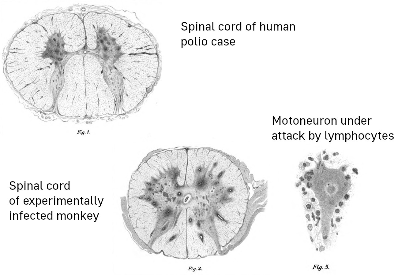
Die Wiener Ärzte Karl Landsteiner und Erwin Popper berichteten die ersten erfolgreichen Experimente zur Übertragung der Polio-Infektion von Menschen auf Versuchstiere, nämlich zwei Affen. Die Tiere entwickelten die typischen Lähmungen, und die pathologischen Veränderungen am Zentralnervensystem entsprachen ebenfalls dem vom Menschen bekannten Bild.

Tolzin und Engelbrecht spekulieren, dass Landsteiners und Poppers Beobachtungen auf nichts weiter beruhen als auf einem unspezifisch krankmachenden Effekt der Injektion von zermahlenem menschlichen Rückenmark in die Bauchhöhle der Versuchstiere. Gegen diese Hypothese sprechen die folgenden Befunde: Die Erkrankung

  • glich in ihren klinischen und pathologischen Manifestationen der Poliomyelitis beim Menschen;
  • manifestierte sich nur bei Affen, aber nicht bei Kaninchen, Meerschweinchen und Mäusen;
  • betraf nur das Zentralnervensystem (insbesondere das Rückenmark), während die inneren Organe unauffällig blieben;
  • blieb ganz aus bei einem dritten Affen, dem man das Rückenmark eines der ersten zwei Affen injizierte. Dieser „Spender“ hatte den Höhepunkt der Erkrankung bereits überschritten, und sein Immunsystem hatte daher das Virus wohl bereits unter Kontrolle gebracht.

Tolzin und Engelbrecht bringen weiterhin vor, dass

die „Pampe“, die Landsteiner und Popper injizierten, beim besten Willen nicht als isoliertes Virus bezeichnet werden kann. Die Forscher legten die genauen Zutaten nicht einmal offen.

Zu den „genauen Zutaten“: Landsteiner und Popper beschreiben ihr infektiöses Material als „mit steriler Kochsalzlösung verriebene Rückenmarkssubstanz in nicht geringer Menge.“ Die Zusammensetzung ist also prinzipiell klar, die genaue Menge allerdings nicht. Immerhin war die resultierende Suspension dünnflüssig genug für eine Injektion, also wohl keine „Pampe“.

Zur Isolation des Virus: Landsteiner und Popper behaupteten gar nicht, das Virus isoliert zu haben. Ihre Schlussfolgerung ist durchaus vorsichtig formuliert:

Da das Rückenmark unserer Fälle sich als sehr infektiös erwies, ohne dass darin Bakterien nachgewiesen werden konnten, ist daran zu denken, dass der Erreger der Poliomyelitis kein durch Bakterienfärbung nachweisbares und nach den bekannten Methoden kultivierbares Virus sei …

Auch diese Studie ist dem interessierten Leser frei zugänglich [5]. Er kann sich also selbst davon überzeugen, dass die Autoren bei ihren Versuchen und bei der Interpretation die angemessene Sorgfalt walten ließen.

Der natürliche Infektionsweg von Polio ist fäkal-oral, d.h. das Virus vermehrt sich im Darmtrakt und wird mit dem Stuhl ausgeschieden. Die unbemerkte orale Aufnahme geringfügiger Mengen davon führt dann beim Empfänger ebenfalls zu einer Darminfektion, mit möglicher Streuung auf dem Blutweg in das Zentralnervensystem. Es ist daher von Interesse, dass eine orale Infektion ebenfalls in Tierexperimenten demonstriert wurde; siehe z.B. Shen et al. [6] und die dort zitierten weiteren Arbeiten. In einer früheren, ausführlicheren Version ihrer Kritik an meinem Artikel haben Tolzin und Engelbrecht die Gültigkeit dieser Studien bestritten, allerdings ohne substanzielle Argumente.

„Nachweis des Poliovirus nicht existent“

Unter dieser Überschrift unterstellen Tolzin und Engelbrecht mir, ich hätte meine Feststellung, dass in der Mehrzahl der klinisch typischen Fälle von Polio das Virus isoliert werden könne, „nicht faktisch untermauern“ können.

Es gibt solche Beweise aber durchaus. McCarroll et al. [3] wiesen das Virus bei einem lokalen Ausbruch in New York in allen vier beobachteten Fällen der paralytischen Erkrankung nach. In einer weiteren Fallserie fanden Bhatt et al. [2] das Virus bei 26 von 31 paralytischen Patienten. In beiden Studien wurde das Virus außerdem auch bei zahlreichen Kontaktpersonen nachgewiesen. Die Erfolgsrate des Virus-Nachweises wird natürlich davon abhängen, wie gründlich und sorgfältig das betreffende diagnostische Labor arbeitet, sowie davon, ob die Proben rechtzeitig entnommen wurden; man kann also nicht unbedingt in jeder berichteten Fallserie solche hohen Nachweisraten erwarten.

Zur grundsätzlichen Frage, welche Methoden zum Nachweis von Viren geeignet seien, habe ich mich anderswo ausführlich geäußert [7]. Dort begründe ich, dass Zellkulturen ein geeignetes und zulässiges Hilfsmittel sind. Dasselbe gilt auch für molekularbiologische Nachweistechniken wie PCR, wenn sie denn mit Umsicht und Sorgfalt eingesetzt werden. Ich muss diese Argumente hier nicht wiederholen.

Zum Nachweis der Wirksamkeit der Impfstoffe

Die aussagefähigsten Untersuchungen zur Wirksamkeit der Polio-Impfung stammen aus der Sowjetunion. Dort wurde, beginnend Mitte 1959, der Sabin-Lebendimpfstoff in großem Stil eingesetzt. Bis Ende 1960 waren 260 Millionen Dosen des Impfstoffs an 77 Millionen Menschen ausgegeben worden [8]. Die Ergebnisse sind in meinem Artikel [1] in Abschnitt 4 zusammengefasst. Die Anzahl der Polio-Erkrankungen ging sofort steil herunter. Sie fiel innerhalb weniger Jahre auf nahe null und blieb dann dort. Derselbe Effekt wurde kurz darauf auch in Ost- und dann in Westdeutschland beobachtet (s.u.).

Tolzin und Engelbrecht schreiben:

Abgesehen davon hätte ein nüchterner Blick auf die Statistiken genügt, um zu erkennen, dass es keinen Grund gab, Salks Impfstoff als den großen Bezwinger eines angeblichen Poliovirus zu feiern. Hier wirkt Palmers Argumentation besonders fragwürdig. So würden einem sicherlich zuerst die historischen Daten aus Ländern wie den USA oder Westeuropas einfallen, wenn es darum geht zu überprüfen, ob die Polioimpfung wirksam war oder nicht …

Hierzu ist zu sagen:

  • Ich habe den Salk-Impfstoff nirgendwo „gefeiert“. Die sowjetischen Studien zeigten eindeutig, dass der von Sabin entwickelte Lebendimpfstoff dem Salk-Totimpfstoff deutlich überlegen war. Nicht ohne Grund wurde kurze Zeit später auch im Westen der Tot- durch den Lebendimpfstoff ersetzt.
  • Die sowjetischen Studien sind besonders aussagefähig, weil sie wirklich generalstabsmäßig durchgeführt wurden – auf eine Weise, wie sie damals im Westen gar nicht möglich gewesen wäre.
  • Bei Impfstudien aus dem Westen unterstellen Kritiker gerne, dass die Ergebnisse durch Einflussnahme von Seiten der Pharma-Industrie verzerrt wurden. Es ist daher etwas seltsam, wenn Tolzin und Engelbrecht jetzt plötzlich westliche Studien zum Maß aller Dinge erheben wollen.

In Wirklichkeit wollen sie aber wohl bloß den Leser von echter, substanzieller Evidenz ablenken und ihn für ihren nächsten Coup in die richtige Stimmung bringen. Dieser besteht in der irreführenden Interpretation einer Statistik aus dem damaligen Westdeutschland. Im Jahr 1961 waren dort 4673 Polio-Fälle gemeldet worden. 1962 wurde die Schluckimpfung eingeführt; im selben Jahr wurden dann nur noch 296 Fälle berichtet.

Um diesen wahrscheinlichen Effekt der Impfung wegzuerklären, bemühen Tolzin und Engelbrecht das ebenfalls 1962 eingeführte Bundesseuchengesetz. Sie präsentieren einen Graphen, welcher zeigt, dass ab demselben Jahr die Zahl der gemeldeten Fälle von Meningitis und Enzephalitis stark anstieg. Sie schließen daraus, dass aufgrund des neuen Gesetzes Krankheitsfälle, die vorher als Poliomyelitis diagnostiziert und gemeldet worden waren, nunmehr als Meningitis oder Enzephalitis gemeldet wurden („Diagnoseverschiebung“). Diese Interpretation ist unhaltbar, aus den folgenden Gründen:

  1. Das neue Gesetz führte zwar eine Meldepflicht für Meningitis ein; Polio blieb aber ebenfalls gesondert meldepflichtig und wurde nicht unter die Meningitis-Kategorie subsumiert.
  2. Eine Infektion mit dem Poliovirus kann zwar als uncharakteristische Meningitis verlaufen und dann möglicherweise unerkannt blieben, aber kein Arzt wird eine klinisch typische Poliomyelitis mit Lähmungen lediglich als Meningitis diagnostizieren.
  3. Bekanntlich galt das Bundesseuchengesetz nicht auf dem Staatsgebiet der DDR. Dort hatte man die Schluckimpfung bereits im Jahr 1960 begonnen, d.h. ein Jahr später als die Sowjetunion, aber zwei Jahre vor der Bundesrepublik. Die Polio-Morbidität ging auch hier schlagartig zurück und betrug im Jahr 1961 nur etwa ein Zehntel des Wertes in der Bundesrepublik [9].

Insgesamt ist festzuhalten, dass die Sowjetunion, die DDR und die Bundesrepublik die Schluckimpfung jeweils in verschiedenen Jahren adoptierten (1959, 1960 und 1962); und in jedem Fall fiel die Zahl der Erkrankung in unmittelbarem zeitlichen Zusammenhang steil ab. Die Wirksamkeit des Lebendimpfstoffs ist also durch substanzielle empirische Daten gut belegt. Nichtsdestoweniger ist dieser Impfstoff – ebenso wie auch der Salk-Impfstoff – mit einer Reihe von Problemen behaftet. Ich spreche diese in meinem Artikel ebenfalls an [1], und ich treffe keine Aussage zu der Frage, ob denn in toto – d.h. im Hinblick nicht nur auf Polio, sondern auf die Gesundheit der Menschen insgesamt – der Nutzen der Impfstoffe ihre Probleme überwiegt.

Industriegifte und Polio

Tolzin und Engelbrecht holen natürlich auch die angebliche Verursachung von Polio durch DDT wieder aus der Mottenkiste. Ich habe meine Argumente gegen DDT als eine wesentliche Ursache von Polio in meinem Artikel dargelegt [1]:

  • In der Sowjetunion ging Polio nach Einführung der Schluckimpfung sofort drastisch zurück, obwohl DDT noch für ein weiteres Jahrzehnt auf annähernd konstantem Niveau eingesetzt wurde.
  • In der Ukraine wurde DDT in sehr viel höherer Dichte eingesetzt als in allen anderen Regionen der Sowjetunion; dennoch waren die Polio-Raten in der Ukraine nicht höher als im Durchschnitt des Landes.

Tolzin und Engelbrecht gehen auf diese Evidenz überhaupt nicht ein. Sie halten mir stattdessen eine Täuschung der Leser vor:

So lässt Palmer unerwähnt, dass sich die erste Anhäufung von Poliofällen 1887 in Schweden ereignete – 13 Jahre nach der Erfindung des Nervengiftes DDT in Deutschland.

Dabei unterschlagen sie selbst allerdings, dass die Anwendung von DDT als Insektizid erst in den 1940er Jahren begann! Der von ihnen konstruierte zeitliche Zusammenhang ist also völlig irrelevant. Zudem schreibt bereits Heine in seiner Polio-Monographie von 1860 [10], ein gewisser Colmann habe über ein epidemisches Auftreten von Polio berichtet. Und wie von Tolzin und Engelbrecht eingangs erwähnt, gibt es durchaus Hinweise auf das Auftreten von Polio bereits im Altertum und im Mittelalter, was bei einer natürlichen Infektionskrankheit ja auch gar nicht anders zu erwarten ist. Literaturhinweise sind in meinem Artikel zu finden.

Ein Zusammenhang von Polio mit DDT lässt sich also mit Sicherheit nicht verallgemeinern. Aber unsere beiden Experten haben natürlich noch weitere Gift-Pfeile im Köcher, wie zum Beispiel Blei und Arsen. Und natürlich gibt es immer noch ein anderes Gift, auf das man ausweichen kann, und noch eines … es wäre ein aussichtsloses Unterfangen, Tolzin und Engelbrecht auf all diesen Fluchtwegen nachzustellen. Natürlich können tatsächlich viele Gifte auch das Nervensystem schädigen. Der zeitliche Verlauf und das klinische Bild werden aber in der Regel von typischer Poliomyelitis abweichen; und natürlich lassen sich solche Gifte auch nachweisen – ebenso wie Polioviren.

Im weiteren verwenden Tolzin und Engelbrecht auch noch viele Worte darauf, mir einen Fehler beim Zitieren eines gewissen Dr. Biskind unter die Nase zu reiben. Ich wurde auf diesen Fehler bereits von anderer Seite hingewiesen und habe meinen Artikel daraufhin auch prompt korrigiert. Diese Korrektur erwähnen meine beiden Kritiker allerdings nicht – die Gelegenheit, mir am Zeug zu flicken, war wohl allzu verlockend.

Fazit

Die Kritik von Tolzin und Engelbrecht enthält nichts Neues. Wie bei Leuten aus dem “No-Virus”-Lager üblich, wählen sie Studien und Fakten auf tendenziöse Weise aus. Diese Auswahl unterziehen sie dann einer ebenso tendenziösen Interpretation; alles wird solange verdreht und verzerrt, bis es zur vorgefassten und gefestigten Ideologie passt.

Eklatant ist dabei auch ihr Doppelstandard für die Bewertung jedweder Evidenz: kein virologischer Befund kann je ihren hohen Ansprüchen genügen, aber andererseits akzeptieren sie vorbehaltlos jede ihrer eigenen Spekulationen als völlig hinreichenden Beweis. Mit solchen Methoden kann man beweisen, was immer man möchte – aber natürlich ist damit jedes Bemühen um echtes wissenschaftliches Verständnis zum Scheitern verurteilt. Tolzin und Engelbrecht haben uns dafür ein anschauliches Beispiel gegeben.

Literatur

  1. (2025) Poliomyelitis und Polioimpfung: eine Kritik an zwei impfkritischen Büchern.
  2. Bhatt, P.N. et al. (1955) Extent of infection with poliomyelitis virus in household associates of clinical cases as determined serologically and by virus isolation using tissue culture methods. Am. J. Hyg. 61:287-301
  3. McCarroll, J.R. et al. (1955) Spread of poliomyelitis infection in nursery schools. Am. J. Public Health Nations Health 45:1541-50
  4. Wickman, I. (1907) Beiträge zur Kenntnis der Heine-Medinschen Krankheit (Karger).
  5. Landsteiner, K. und Popper, E. (1909) Übertragung der Poliomyelitis acuta auf Affen. Z. Immunitätsforsch. 2:377-393
  6. Shen, L. et al. (2017) Pathogenic Events in a Nonhuman Primate Model of Oral Poliovirus Infection Leading to Paralytic Poliomyelitis. J. Virol. 91
  7. Palmer, M. (2024) Der Irrweg der “`No Virus”‘-Doktrin.
  8. Chumakov, M.P. et al. (1961) Some results of the work on mass immunization in the Soviet Union with live poliovirus vaccine prepared from Sabin strains. Bull. World Health Organ. 25:79-91
  9. Lindner, U. (ed.: Schulze, H.) (2004) Gesundheitspolitik in der Nachkriegszeit — Großbritannien und die Bundesrepublik Deutschland im Vergleich (German Historical Institute London).
  10. von Heine, J. (1860) Spinale Kinderlähmung (Cotta).

Poliomyelitis and polio vaccination: a critique of two vaccine-critical books

Poliomyelitis and polio vaccination: a critique of two vaccine-critical books

Michael Palmer, MD

1. Two books on the safety and efficacy of childhood vaccines that are worth reading

no image info

Readers may be familiar with these two books [1,2]. “Dissolving Illusions” has recently been updated and expanded, but this critique is based on the first edition from 2013.

Both books provide valuable information. I particularly appreciate “Dissolving Illusions” for its rich historic perspective, whereas “Turtles” thoroughly documents the astonishing lack, seemingly pertaining to all vaccines, of properly conducted studies to show their efficacy and safety. However, with both books, I found the treatment of poliomyelitis unconvincing, which prompted me to give this subject a closer look. This paper is the result.

2. What causes poliomyelitis?

2.1. Poliomyelitis according the textbooks

no image info
  • Oral viral infection
  • Multiplication in the gut mucosa, fecal excretion
  • Spinal cord infection with paralytic disease in a small percentage of cases

According to the the mainstream view, polio is caused by a viral infection. There are three strains of poliovirus, all of which belong to the enterovirus family, like for example hepatitis A and Coxsackie viruses. One of the three strains is now considered extinct.

As with many other enteroviruses, the infection occurs through oral uptake, whereupon the virus multiplies in the gut. It is then excreted in the stool, and fecal contamination leads to oral uptake by the next person. Such fecal-oral transmission is a common pattern with many other enteric pathogens also.

In the the majority of infected persons, symptoms will be limited to mild, uncharacteristic and self-limiting enteric disease. Some others can have viral meningitis, manifest as headache, high fever and so on, but still no paralytic disease.

Only rarely will the CNS itself be affected, and in particular the motoneurons in the anterior horns of the spinal cord and the brainstem. Destruction of these cells will disconnect the muscle cells under their control, and flaccid paralysis will be the result. Paralysis is observed only in about one in a thousand cases. Depending on the extent of the destruction, the paralysis can be transient or permanent, and in some cases deadly. It is not uncommon for paralytic patients to recover some but not all of their motor control.

2.2. Poliomyelitis according to “Turtles”

  • No clear evidence that poliomyelitis is caused by poliovirus
  • Assumes higher polio incidence
    • in the 20th than in the 19th century
    • in developed countries than in developing ones

    … and interprets this as contradicting the conventional narrative of the poliovirus

  • Proposes DDT and other pesticides as the real causes of the disease
  • Treats polio vaccine efficacy as doubtful, but bases this verdict on early findings with the Salk (dead) vaccine alone, does not discuss the Sabin live vaccine
  • Harps on the “Cutter incident”, without understanding the implications
  • Confused discussion of the role of enteroviruses other than poliovirus in paralytic disease

While both books are highly critical of the conventional polio narrative, “Turtles” is the more radical of the two, proposing that poliomyelitis has nothing to do with the poliovirus and is instead caused by DDT and possibly by other environmental poisons. To make its case, it emphasizes epidemiological “enigmas” such as the supposed association of poliomyelitis with modern western civilization. In its zeal to malign polio vaccination, it harps on the “Cutter incident”, which involved the occurrence of poliomyelitis in recipients of an early batch of the Salk vaccine, apparently unaware that this unfortunate event strongly corroborates the viral causation of the disease.

2.3. Poliomyelitis according to “Dissolving Illusions”

  • Broadly similar to “Turtles” (but predates it)
  • Does not dispute that poliovirus can cause poliomyelitis, yet still emphasizes the role of DDT and other factors:
Refined sugar, white flour, alcohol, tobacco, tonsillectomies, vaccines, antibiotics, DDT, and arsenic … Many thousands of people were needlessly paralyzed because the medical system refused to look at the consequences of these …

“Dissolving Illusions” is less radical, but this leaves the reader even more confused as to the question of disease causation. On the one hand, the book appears to accept that polioviruses can induce it, yet on the other it suggests that the sinfulness and debauchery of modern civilization are the true underlying causes—or maybe alternate ones; this distinction is not clearly addressed.

3. What evidence indicates that poliomyelitis is caused by viral infection?

  • Infectious nature meticulously documented by Ivar Wickman (Swedish polio epidemic of 1905)—transmission mostly through people with abortive (non-paralytic) disease
  • Experimental transmission of the infection from deceased child to two monkeys (Landsteiner and Popper, 1909)
  • Experimental transmission via oral route to monkeys shown in numerous later studies
  • Poliovirus can be isolated in the majority of clinically typical cases (and from the CNS of fatal cases)
  • Efficacy of vaccination (particularly the live vaccine)

A transmissible nature of the disease had been suggested by earlier authors, but convincing evidence was compiled for the first time by the Swedish pediatrician Ivar Wickman [3]. His meticulous study of about 1000 cases of poliomyelitis that had occurred during the Swedish epidemic of 1905 showed convincingly that paralytic poliomyelitis was only the most severe form of the infection. Most cases were in fact “abortive”, i.e. clinically non-characteristic and indistinguishable from gastroenteritis or viral meningitis due to other causes. Nevertheless, even very mild cases could still transmit the infection to others, in whom paralytic disease might then become manifest.

Wickman’s diligence also shines through in his attempts to isolate a causative bacterium—unlike some contemporaries, he did not find one, and he concluded that an unknown virus must be the cause. This was soon confirmed by other investigators.

3.1. Landsteiner and Popper’s experiment

no image info

Wickman’s findings prompted the Viennese researchers Karl Landsteiner and Erwin Popper to attempt the experimental infection of monkeys, using CNS tissue samples from a boy who had succumbed to the disease. Both monkeys injected with these samples developed paralytic disease, and their CNS lesions closely resembled those from human victims. The attempt to infect further animals with tissue samples from the second monkey failed, most likely because the latter had been let live long enough to start producing neutralizing antibodies or even to extinguish the infection altogether. However, other researchers soon established serial passage in monkeys and subsequently other animals.

“Turtles” maintains that these early findings are invalid, because they relied on injection instead of on the natural oral route of infection. However, the book ignores numerous later studies that induced poliomyelitis in monkeys after oral infection; only some examples are cited here for illustration [4–7].

The drawings in this slide are taken from Landsteiner and Popper’s original study [8]. The dark splotches in the two spinal cord cross-sections are hemorrhagic foci of infection. The magnified view of a single large nerve cell surrounded by immune cells captures the pathogenetic mechanism of the disease: virus-infected motor nerve cells are destroyed by an immune attack, which results in the paralysis of the muscle fibers controlled by them.

At the time of these experiments, there were no methods for actually purifying and characterizing virus particles. Later technical advancements led to the isolation of three immunologically distinct poliovirus strains, all of which belonged to the enterovirus family. Both the Salk (inactivated) and the Sabin (live) vaccine contain derivatives of all three strains.1

4. Efficacy of polio vaccination: the Russian experience

  • The Soviet Union adopted the Sabin live vaccine in 1959
  • Some 15 million persons received oral vaccine in 1959, and 77.5 million in 1960 (mainly between 2 months and 20 years old)
  • For the sake of comparison, some administrative districts within the USSR received the inactivated (Salk) vaccine instead

Even though both the inactivated (“dead”) and the live polio vaccines were invented in America, the first country to test the live vaccine on a large scale was the USSR. The results, and the major logistics effort involved, are described in a study by Chumakov [9].

The paper acknowledges the support by Albert Sabin (the inventor of the live vaccine) and discusses the reason for going “all-in” at once: by vaccinating most of the susceptible population in one fell swoop, the risk would be minimized that the vaccine virus strains could spread serially from one susceptible person to the next, and in the process might acquire mutations that would restore their neurovirulence. It has since been found that this concern was well-founded; less thorough and comprehensive vaccination campaigns have indeed given rise to such mutations (see e.g. [10]).

4.1. Acute polio cases per year in the USSR before and after vaccination

no image info

The outcome of the vaccination campaign that was begun halfway through the year 1959 is summarized in this graph from a later paper [11]: the annual incidence of the disease dropped sharply and stayed down during the two subsequent decades.

Considering the very high vaccination rate achieved already in 1960, one might wonder why the initial drop was not even steeper than apparent from this slide. While this question cannot definitely be answered from the studies at hand [9,11], this may have to do with increasingly strict diagnostic criteria: cases may have initially been diagnosed based on clinical findings alone, whereas in later years only cases confirmed by detection of the virus or specific antibodies may have been counted. Alternatively, the vaccine strains might have with time dislodged the circulating wild type poliovirus strains. A definite answer would likely require a thorough examination of the Russian medical literature.

4.2. Efficacy of live vs. inactivated polio vaccine

no image info

While most young persons in Russia had received the live vaccine, an inactivated vaccine was used for comparison in some administrative districts. The graph in this slide (from data given in [9]) shows that residual rates of polio remained distinctly higher in those districts, showing a clearly inferior efficacy of the inactivated vaccine. In fact, in the Krasnodar district, the case numbers in 1960 exceeded the national averages from the years before the vaccination campaign.

The lower (or even marginal) protective efficacy of the inactivated vaccine may have been due to excessive chemical treatment. At the time of this campaign, the Cutter incident in the U.S. had already occurred—an incompletely inactivated vaccine had given rise to paralytic disease in several hundred vaccinees. The Russian scientists may have erred on the side of caution by applying too aggressive chemical inactivation. This would have “mangled” the immunological features of the virus particles and rendered any antibodies induced by them largely ineffective against the unmodified virus.

Production methods for the inactivated vaccine have since improved, and other countries that have relied on this vaccine have obtained better results [12]. Nevertheless, the data in this slide show that “Turtles” and “Dissolving Illusions” are remiss in basing their evaluation of polio vaccine efficacy only on experiences with early inactivated vaccines from the 1950s.

5. Serious problems with polio vaccines

  • Failed immunization or fading immunity
  • Persons who have received the inactivated vaccine may still spread the wild-type virus
  • Live vaccine virus may cause paralytic disease
  • Inactivated vaccine may contain residual live virus, giving rise to paralytic disease
  • Early batches of both live and inactivated vaccine were contaminated with the oncogenic monkey virus SV40

While though I consider the evidence of vaccine efficacy against polio conclusive, I do not mean to imply that there are no serious problems with the vaccines. In fact, I consider it unproven that any of these vaccines have produced a net benefit in overall health outcomes.

Failed immunization or fading immunity are more common with the inactivated vaccine, but infections with wild-type poliovirus strains leading to paralytic disease have occurred in live-vaccinated persons also. In principle, fading immunity can be overcome with booster injections, but such campaigns tend to have lower participation rates. This can leave sizable numbers of people without immunity and result in outbreaks of the wild-type polio.

One such outbreak occurred in Albania in the year 1996; it was reportedly stopped by subsequent mass vaccinations. In this outbreak, the age groups between 11 years and 35 years were preferentially affected [13]. It seems that these age groups had previously received vaccine that had been stored without refrigeration for several weeks.

5.1. Live virus vaccine and paralytic disease

  • The live virus strains are attenuated, but in immunocompromised persons they may nevertheless cause paralytic disease (numerous cases reported, some fatal)
  • Live vaccine virus strains may be transmitted from vaccinated to unvaccinated persons, including immunodeficient ones
  • Serial transmission of vaccine virus can lead to mutations that give rise to increased neurovirulence
  • There have been multiple outbreaks of poliomyelitis due to mutated vaccine virus strains

Even though the live vaccine virus strains are less virulent than the wild-type forms, a functioning immune system is nevertheless required to contain the infection with these vaccine viruses. In persons with genetic or acquired immune deficiencies, these attenuated strains can cause paralytic disease, sometimes fatal.

Immunocompromised people may also have difficulty clearing the infection. The prolonged replication in such persons gives the vaccine virus time to mutate and thereby revert to increased virulence. Such mutated strains may then be transmitted to the non-immune population at large. Multiple outbreaks due to mutant vaccine virus strains have indeed occurred [10,14,15]. Conceivably, such strains might actually perpetuate polio after a successful eradication of the wild-type strains.

5.2. SV40 contamination of early polio vaccine batches

From Fields Virology:

The next member of the [polyomavirus] family to be isolated was simian virus 40 (SV40) by Sweet and Hilleman in 1960. They were screening samples from poliovirus vaccine lots produced in rhesus monkey kidney cells for the presence of contaminating viruses. SV40 was the 40thvirus isolated in this screen …

From Sweet and Hilleman’s paper:

Hull et al. have studied extensively viruses encountered in “normal” monkey renal cell cultures. Hull called them simian or SV viruses … 28 of these viruses were precisely separated serologically into types and, additionally, 24 unidentified viruses were recorded.

… other viruses may also have been in some vaccine batches


The polyomavirus SV40 was present as a contaminant in early batches of both the live and the inactivated polio vaccines. It came from the monkey kidney cells that were used to grow up those vaccines. The standard text “Fields Virology” [16] offers a somewhat inaccurate version of these events, but the paper by Sweet and Hilleman it cites sets the record straight [17]. Even before these authors got involved, a large number of contaminating monkey viruses had been identified by Hull and coworkers.

While it seems that all of these scientists tried their best to prevent the release of contaminated vaccine batches, they had to rely, for the detection of so far unknown viral agents, on observing cytopathic effects in cell culture, i.e. of infected cells that look sick or die off. It seems that SV40 did not readily produce such cytopathic effects in the cell cultures used for screening those vaccine lots.

Sweet and Hilleman reported the repeated isolation of SV40 as early as 1960, yet nevertheless the virus remained in commercial vaccine batches for several years after that. It seems that this was covered up by the authorities.

5.3. SV40 and human health

  • SV40 induced tumors in animal experiments
  • Has been detected in various kinds of human tumors
    • mesothelioma
    • brain cancer
    • bone cancer
    • non-Hodgkin-lymphoma
  • Statistical studies disagree on the correlation between polio vaccines and cancer
  • Heinonen 1973 [18] found low absolute case numbers, but high relative risk of cancer among children exposed to inactivated vaccine in in utero

The association of SV40 with some human tumors seems to be uncontroversial, but some may still question the causal role of the virus in these tumors [19]. It is unclear to what extent SV40 in the polio vaccines has increased the risk of such cancers. Most statistical studies find no clear evidence of cancer risk due to these vaccines [20].

One study stands out, however, with respect to both its findings and the group of subjects investigated. Heinonen et al. [18] examined the overall risk of cancers in children who had been exposed in utero, i.e. their mothers had been injected with the inactivated vaccine during pregnancy. While the absolute case numbers of cancers in those children were not particularly high, the relative risk was significantly increased.

In this connection, we should note that other polyomaviruses are known to cause disease in immunocompromised patients. The JC virus causes progressive multifocal leukencephalopathy, a form of wholesale brain degeneration, whereas the BK virus causes a very destructive disease of the kidneys. Embryos and early fetuses, too, lack a functioning immune system; this makes them susceptible for example to severe damage by rubella virus, which after birth will only very rarely cause major disease. It is not implausible to assume that exposure to SV40 in utero may cause more widespread infection that, even though not manifesting as dramatically as rubella, may then give rise to cancer sometime after birth.

We might also wonder whether SV40 would be more prone to cause disease when contained in live vaccine than in the inactivated vaccine or vice versa. On the one hand, the live vaccine is not subject to chemical inactivation, so that contaminating viruses would more likely survive. On the other hand, since the live vaccine is not injected, such contaminating viruses would have to traverse the intestinal barrier. Thus, the question cannot be answered from a priori considerations. I am not aware of any thorough experimental investigation of this question.

6. DDT and Polio

As stated before, both “Turtles” and “Dissolving Illusions” maintain that, in its day, DDT was a major cause of epidemic poliomyelitis. We can use the Russian data to evaluate this claim.

6.1. DDT use in the USSR by region

no image info

In the USSR, DDT was mostly used in agriculture. This graphic, taken from [21], shows the cumulative amounts used per unit area over the middle decades of the 20thcentury. Ukraine, when part of the former USSR, used to be that country’s richest and most fertile region, and accordingly this is where DDT was used the most extensively.

6.2. Polio in the Ukraine vs. USSR overall

no image info

In keeping with the high amounts of DDT used in Ukraine, we should expect a higher incidence of polio in this area than in the remainder of the Soviet Union (and therefore than the national average). However, this slide shows that both before and after the onset of vaccination, the incidence in Ukraine closely tracked the national average [9].

6.3. Time course of polio incidence and of DDT use in the USSR

no image info

That DDT was not the cause or a major contributing factor to poliomyelitis is also evident from this slide, which compares the time course of polio incidence [11] to that of DDT use [21] in the Soviet Union.

Polio rates dropped precipitously immediately after the onset of vaccination, whereas DDT use continued on a high level for another decade before being phased out. When its use was finally terminated, this had no discernible effect on the already very low incidence of poliomyelitis.

6.4. “Dr. Biskind goes into battle”

“Turtles” quotes Dr. Morton Biskind to prop up its own story about DDT causing polio:

During a period of more than two years, numerous cases of a curious symptom complex, apparently never before reported … widely attributed to infection with a thus far illusory “virus.” [22]
The high incidence, the usual absence of a febrile reaction … suggested an intoxication rather than an infection … soon led to consideration of DDT. [23]

The title of this slide is taken directly from “Turtles.” Thus, the authors appoint Dr. Morton Biskind, posthumously, as the champion of their failed DDT hypothesis. However, a closer look at Biskind’s statements shows that he had implied nothing of the sort.

6.5. “Dissolving Illusions” and its interpretation of Biskind’s words

In the fear-baked summers of polio, many parents were totally unaware that exposure to DDT alone induced symptoms that were completely indistinguishable from poliomyelitis—even in the absence of a virus.

6.6. What is wrong with this picture?

  • Biskind cites a combination of symptoms “never before reported”—but polio is clinically familiar
  • Polio typically begins with a febrile infection, usually involving the GI tract (vomiting/diarrhea) and the meninges (headache etc.)—Biskind’s words “absence of a febrile reaction” do not describe polio
  • The new “virus X” had been postulated precisely because these cases did not resemble polio or any other familiar infectious disease
  • Biskind’s 1949 paper mentions poliomyelitis only once, in a footnote:
Needless to say, findings related to the nervous system, and muscular spasm and weakness in severe acute affections of this type have led to confusion with such entities as meningitis and poliomyelitis.

Biskind is clearly saying that DDT likely caused numerous cases of intoxication, which were mistaken by his colleagues not for polio but for a novel infectious disease. Only a few individual cases were conflated with familiar diseases such as meningitis or polio. Errors of this kind are always bound to happen, but Biskind accords them no more than a footnote.

“Dissolving Illusions” commits the same blunder as “Turtles”, citing Biskind’s 1949 paper as evidence for the following statement:

In the fear-baked summers of polio, many parents were totally unaware that exposure to DDT alone induced symptoms that were completely indistinguishable from poliomyelitis—even in the absence of a virus.

The meaning of Biskind’s words is clear enough, and it has no similarity to that which “Turtles” and “Dissolving Illusions” impute to them.

Correction: Zoey O’Toole, the translator and editor of “Turtles”, has pointed out to me that in some later papers Dr. Biskind indeed stated more clearly his suspicion that cases of DDT poisoning had been mistaken for polio:

When the population is exposed to a chemical agent [DDT] known to produce in animals lesions in the spinal cord resembling those in human polio, and thereafter the latter disease increases sharply in incidence and maintains its epidemic character year after year, is it unreasonable to suspect an etiologic relationship?

“Turtles” indeed cites the paper containing this statement, whereas “Dissolving Illusions” does not. While this correction invalidates my criticism regarding the use of Dr. Biskind as a witness, it does not validate the books’ scientific arguments.

7. Paralytic disease related to polio

Both books discuss poliomyelitis and clinically similar paralytic diseases, suggesting that such diseases somehow invalidate the conventional polio narrative. However, these insinuations are without foundation.

7.1. Poliomyelitis without poliovirus

  • Several enteroviruses other than poliovirus have been shown to cause the same disease and lesions in the spinal cord
  • Usually clinically milder than with poliovirus, but enterovirus 71 has proven highly neurovirulent in some outbreaks
  • “Nonparalytic poliomyelitis” is not clinically characteristic—confusion on this point arises from confused nomenclature

The entire subject was lucidly reviewed by Sabin in 1981 [24], but neither book cites this outstanding paper. Sabin enumerates the enteroviruses that had at the time been found to cause poliomyelitis. Individual cases of this disease were clinically and pathologically indistinguishable from those caused by the “proper” poliovirus strains, but on average their clinical course tended to be milder.

In assessing the relative importance of these virus strains, one must take into account that polio vaccination has greatly reduced the incidence of disease caused by poliovirus, whereas no protection is available from these more recently discovered viruses. Thus, a significant contribution of novel viruses to overall disease remaining after the introduction of polio vaccines does not detract from the usefulness of those vaccines.

Our two books also try to make hay by pointing to the frequent failure of poliovirus isolation from clinically diagnosed cases of “non-paralytic poliomyelitis.” But this diagnosis is a misnomer—a contradiction in terms. “Poliomyelitis” really refers to a pathological finding, namely, an inflammation of the spinal cord’s gray matter that cannot fail to induce at least transient paralysis.

To avoid such confusion, Wickman had already proposed in 1907 the name “Heine-Medin disease” to comprise both paralytic and non-paralytic infections with the same (at the time still hypothetical) infectious agent [3]. It is unsurprising that, in the context of an ongoing polio epidemic, some cases of gastroenteritis or viral meningitis will initially be attributed to poliovirus, but then turn out to be due to other pathogens instead. The exact percentages of such mistaken clinical diagnoses have of course no bearing whatsoever on the actual significance of poliovirus.

7.2. Paralytic poliomyelitis in spite of vaccination

Several possible causes:

  • Other neuropathogenic viruses (particularly enteroviruses)
  • Failed immunization or fading immunity
  • Other acute neurological diseases can be clinically difficult to differentiate, e.g. polyradiculitis (Guillain-Barré syndrome)

We have already touched on several of these causes. The live vaccine is applied orally and recapitulates the normal gastrointestinal infection, which also means that it is shed in the stool and can be transmitted to other persons, much like the wild-type strains of poliovirus. As noted earlier, this can give rise to mutant vaccine strains that revert to higher virulence. It also poses a problem in connection with immunodeficient individuals, who may become exposed not only by being vaccinated themselves, but also indirectly due to the shedding of vaccine virus by other persons.

Guillain-Barré syndrome (polyradiculitis) can be difficult to distinguish from poliomyelitis, although a combination of virological, immunological and electrophysiological tests (and if necessary nerve biopsies) should resolve most clinically ambiguous cases. The disease is due to cross-reactive autoimmunity that is triggered by natural infections, but also by vaccinations, including those against polio.

If one aims for a valid risk-benefit analysis of polio vaccination, the disease forms listed here must certainly be taken into consideration. Alas, neither “Turtles” nor “Dissolving Illusions” attempt such a balanced evaluation.

7.3. Polio and “acute flaccid paralysis”—globally reported cases 1988-2017

no image info

This chart is shown as Figure 10.3 in “Turtles.” The book comments on the findings as follows:

The number of cases of non-polio acute flaccid paralysis has been steadily rising, in parallel with the decrease in polio incidence.

The subtext is that, since poliovirus (according to “Turtles”) is not the cause of poliomyelitis, “acute flaccid paralysis” (AFP) is merely a new name for the disease formerly known as polio. However, the following observations are in order:

  • The graph merely shows globally reported cases; there is no way of knowing for certain what the incidence of unreported cases of AFP was in earlier years.
  • AFP is a broad clinical category, but individual cases can be more exactly attributed to Guillain-Barré syndrome, true poliomyelitis, and other causes. There is no need to speculate.
  • If we take the graph at face value, then it is clear that AFP took off not “in parallel with the decrease in polio incidence” but only after polio had already dropped to near zero.

As with their DDT story, it is obvious that the authors of “Turtles” cannot see past their own preconceived notions and look at the data without prejudice.

8. Is poliomyelitis really a disease of civilization?

  • In the west, did polio really become prevalent/epidemic only in the 20thcentury?
  • In colonial settings etc., did polio really affect westerners more than the local populations?
  • Are noble savages really protected from poliomyelitis?

The idea of polio as a disease of civilization pervades much of the literature on the disease, and, needless to say, our two books enthusiastically run with it. But does it stand up to scrutiny?

8.1. Findings reported by Jabob Heine in 1860

  • German MD orthopedist, first physician to systematically study the disease
  • Treated 192 poliomyelitis patients in his own clinic, achieving significant clinical improvement in many cases
  • Regarding the prevalence of the disease, he noted (my translation):
[The disease] may properly be called widespread, and not only in our part of the world … such sudden paralysis in small and otherwise healthy children also frequently occurs in India, and we hear similar reports from Egypt and other non-European countries. Another author, Colmann, even mentions that it occurs epidemically … also, almost all paralytic deformations that date from childhood belong to this form of paralysis.

Among the very limited sources on poliomyelitis from the 19thcentury, the monograph “Spinale Kinderlähmung” [25] by Jacob Heine stands out. Heine was the first physician to make this disease his main focus of study, and according to his own reports he had considerable success in treating (and sometimes curing) residual paralysis. His quoted words, as well as the very considerable number of patients in his own care, do not encourage the idea that the disease was less common in his day than 100 years later.

Another case in point is the Swedish epidemic studied by Wickman—even though it had occurred a little later, shortly after the turn of the century. In this epidemic, the incidence had been higher in the rural farming communities than in the cities, again giving no indication that modernity was somehow responsible for spreading the disease.

A more plausible explanation for the perceived increase in importance of polio is the greatly diminished incidence of other childhood diseases—polio came to the fore simply by not being likewise diminished.

8.2. An apparent case of polio on a stele from ancient Egypt

no image info

This Egyptian stele has been dated to about 1500 years B.C. It shows the guardian of the temple of the goddess Astarte, with his wife and son. The atrophy and deformity of his right leg are of course merely suggestive of polio. However, there is a good number of skeletons dating from antiquity and the middle ages with deformities that have been ascribed to polio by the anthropologists who have studied them [26].

8.3. Albert Sabin on polio incidence in China

In 1946, I investigated an outbreak of poliomyelitis among U.S. Marines in Tientsin, China. I was told at the time that paralytic poliomyelitis was extremely rare in China. Some years later, Ku Fang-chou et al. reported a very high incidence of paralytic poliomyelitis in Tientsin, Shanghai, and Tsingtao in 1957-1959 … the average annual rates of reported paralytic cases of 144, 263, and 353 per million total population in these three cities was higher than the ~135 reported paralytic cases per million total population in the United States during the 1951-1955 prevaccine era.

Sabin, a medical man of outstanding ability, dedicated his life’s work to understanding and fighting poliomyelitis. Toward the end of his career, he came to the conclusion that “much of the dogma about paralytic poliomyelitis in the tropics was based on obviously incomplete reporting” [24].

8.4. That leaves the noble savages …

“Dissolving Illusions” cites a study by Neel et al. [27], concerning the isolated Brazilian Xavante tribe:

The paradox of a virtual absence of paralytic poliomyelitis among such heavily infected groups as this, despite high antibody titers, is well known, but the interpretation of the observation remains under discussion.

But how profound is the mystery, really?

  • Neel reports that “there are today between 1500 and 2000 Xavante, living in a number of autonomous communities along the Rio das Mortes …”
  • Assuming the same incidence as in the US before vaccination, there should be about 0.2 to 0.25 new cases per year

Neel also mentions that the study was “carried out during the summer of 1962,” i. e. it lasted less than a year. Therefore, even considering that poliomyelitis is more common in the summer than in the winter—but such seasonality is less pronounced in warmer climates—the above numbers mean that Neel’s chances of witnessing any acute cases at all would have been quite low.

As to the prevalence of persistent paralysis, the vagueness of his population estimate (“between 1500 and 2000”) strongly suggests that he did not make a close first-hand survey of that population, which might have acquainted him with any such cases. Thus, no mystery remains to be explained or explained away.

9. Summary

Both books

  • present important facts and problems with polio vaccines
  • … but weaken their own case by not offering a balanced perspective
  • don’t acknowledge the effectiveness of the live vaccine
  • push a wrong-headed idea of disease causation by DDT, which conforms to naive environmentalist prejudice but does not withstand scrutiny
  • obsess over “epidemiological enigmas” that have no solid basis in fact

While both books present important information on polio vaccines that must be part of any valid risk-benefit analysis, neither offers a balanced, realistic overall perspective. Thus, these books should not be relied upon as the sole sources of information in making decisions for or against polio vaccination.

Notes

  1. Subsequent to the apparent extinction of one of the wild-type strains, bivalent vaccines against the remaining two strains have been introduced. (go back)

References

  1. Humphries, S.A.B.R. (2013) Dissolving Illusions: Diseases, Vaccines, and the Forgotten History (CreateSpace).
  2. Anonymous (2022) Turtles All The Way Down: Vaccine Science and Myth (Children’s Health Defense/Skyhorse Publishing).
  3. Wickman, I. (1907) Beiträge zur Kenntnis der Heine-Medinschen Krankheit (Karger).
  4. Trask, J.D. and Paul, J.R. (1941) Experimental Poliomyelitis In Cercopithecus Aethiops Sabaeus (The Green African Monkey) By Oral And Other Routes. J. Exp. Med. 73:453-9
  5. Horstmann, D.M. et al. (1947) The Susceptibility Of Infant Rhesus Monkeys To Poliomyelitis Virus Administered By Mouth : A Study Of The Distribution Of Virus In The Tissues Of Orally Infected Animals. 99 86:309-23
  6. Howe, H.A. and Bodian, D. (1948) Poliomyelitis in the cynomolgus monkey following oral inoculation. Am. J. Hyg. 48:99-106
  7. Gebhardt, L.P. and Bachtold, J.G. (1953) Reproducible oral infection rates in monkeys with the Mahoney strain (Type 1) of poliomyelitis virus. 431 83:807-9
  8. Landsteiner, K. and Popper, E. (1909) Übertragung der Poliomyelitis acuta auf Affen. Z. Immunitätsforsch. 2:377-393
  9. Chumakov, M.P. (1961) Some results of the work on mass immunization in the Soviet Union with live poliovirus vaccine prepared from Sabin strains. Bull. World Health Organ. 25:79-91
  10. Burns, C.C. et al. (2013) Multiple Independent Emergences of Type 2 Vaccine-Derived Polioviruses during a Large Outbreak in Northern Nigeria. 377 87:4907-4922
  11. Grachev, V.P. (1984) Long-term use of oral poliovirus vaccine from Sabin strains in the Soviet Union. 2861 6 Suppl 2:S321-2
  12. Axelsson, P. (2012) The Cutter incident and the development of a Swedish polio vaccine, 1952-1957. Dynamis 32:311-328
  13. Prevots, D.R. et al. (1998) Outbreak of paralytic poliomyelitis in Albania, 1996: high attack rate among adults and apparent interruption of transmission following nationwide mass vaccination. Clin. Infect. Dis. 26:419-25
  14. Etsano, A. et al. (2016) Environmental Isolation of Circulating Vaccine-Derived Poliovirus After Interruption of Wild Poliovirus Transmission – Nigeria, 2016. MMWR 65:770-3
  15. Korotkova, E.A. et al. (2016) A Cluster of Paralytic Poliomyelitis Cases Due to Transmission of Slightly Diverged Sabin 2 Vaccine Poliovirus. J. Virol. 90:5978-88
  16. Knipe, D.M. and Howley, P.M. (2013) Fields Virology (Wolters Kluwer).
  17. Sweet, B.H. and Hilleman, M.R. (1960) The vacuolating virus, S.V. 40. Proc. Soc. Exp. Biol. Med. 105:420-7
  18. Heinonen, O.P. et al. (1973) Immunization during pregnancy against poliomyelitis and influenza in relation to childhood malignancy. Int. J. Epidemiol. 2:229-35
  19. Ferber, D. (2002) Virology. Monkey virus link to cancer grows stronger. Science 296:1012-5
  20. Dang-Tan, T. et al. (2004) Polio vaccines, Simian Virus 40, and human cancer: the epidemiologic evidence for a causal association. Oncogene 23:6535-40
  21. Li, Y.F. et al. (2006) Dichlorodiphenyltrichloroethane usage in the former Soviet Union. Sci. Total Environ. 357:138-45
  22. Biskind, M.S. (1949) DDT poisoning and elusive virus X; a new cause for gastro-enteritis. Am. J. Dig. Dis. 16:79-84
  23. Biskind, M.S. (1951) Statement on clinical intoxication from DDT and other new insecticides. J Insur Med 1946 6:5-12
  24. Sabin, A.B. (1981) Paralytic poliomyelitis: Old dogmas and new perspectives. Rev. Infect. Dis. 3:543-64
  25. von Heine, J. (1860) Spinale Kinderlähmung (Cotta).
  26. Berner, M. et al. (2021) Challenging definitions and diagnostic approaches for ancient rare diseases: The case of poliomyelitis. Int. J. Paleopathol. 33:113-127
  27. Neel, J.V. et al. (1964) Studies On The Xavante Indians Of The Brazilian Mato Grosso. Am. J. Hum. Genet. 16:52-140
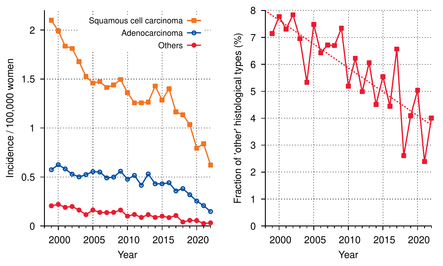
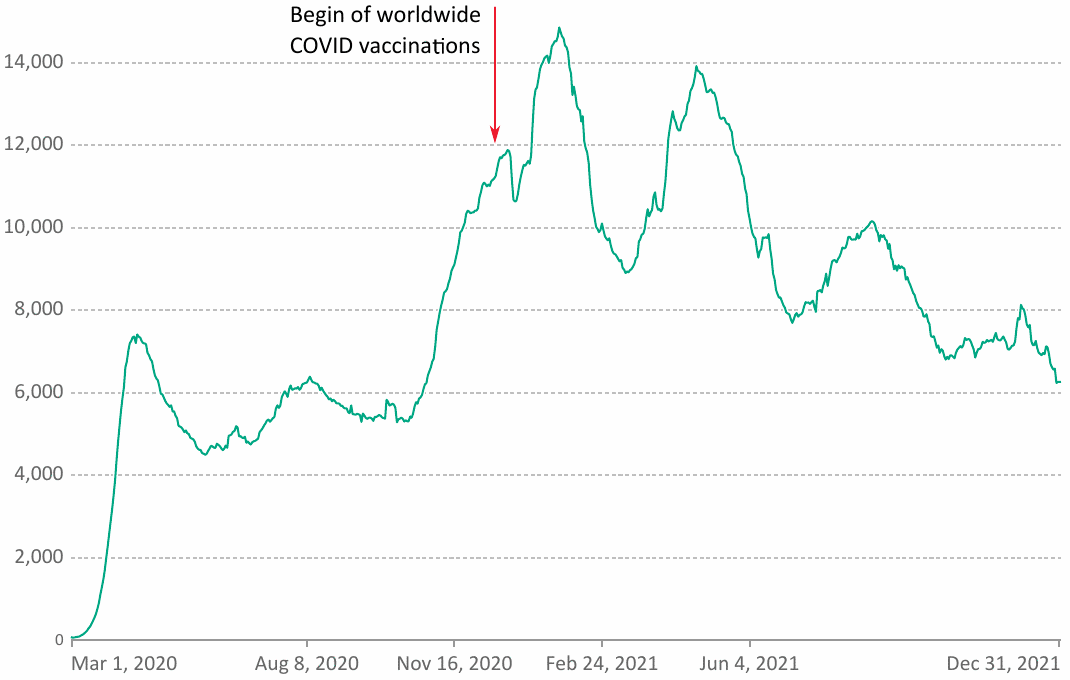
The EU Commission can see no excess mortality due to the COVID-19 vaccines

Michael Palmer, MD and Sucharit Bhakdi, MD

I only believe in statistics that I doctored myself.
Winston Churchill

1. Background

On August 29, 2023, Ivan Vilibor Sinčić, a Croatian member of the EU Parliament, posed the following simple and poignant question to the EU Commission:

Per the European Medicines Agency’s EudraVigilance system, how many people have been reported dead as a consequence (side effect) of receiving approved COVID-19 vaccines since the administration of these medical products began?

On November 6, he received the following written reply from Ms. Stella Kyriakides, the EU Commissioner for Health and Food Safety:

EudraVigilance is a database collecting suspected side effects reported by the European Economic Area’s (EEA) patients and healthcare professionals, i.e. medical events reported following the use of a medicine in the EEA.The fact that these events were observed following the use of the medicine does not mean that they were caused by it. They may have been caused by underlying medical conditions of the individual, by other medicines taken in parallel or due to other events entirely.

Scientific studies investigate potential causal links in these temporal associations and most suspected side effects are not eventually confirmed as side effects.

An unprecedent [sic] high number of people has been administered COVID-19 vaccines [1] and the number of reported suspected side effects is consequently much higher than for other medicines.

As of 30 September 2023, EudraVigilance shows 11,977 spontaneous reports of suspected side effects with reported fatal outcome for all authorised COVID-19 vaccines.

Only in very exceptional cases, deaths have been reported to be caused by the vaccine. One example is ‘thrombosis with thrombocytopenia syndrome’ with adenoviral vector COVID-19 vaccines [2] for which warnings and contraindications have been included in the product information to inform healthcare professionals and patients and reduce risk of adverse consequences.

There is no evidence that COVID-19 vaccines are causing excess mortality [3] and no safety signal for increased mortality with any of the authorised COVID-19 vaccines has been identified by EMA to date. In fact, COVID-19 vaccines have saved millions of lives.

[1] Almost 768 million vaccine doses administered in EU and EEA countries.
[2] https://www.nature.com/articles/s41541-022-00569-8
[3] https://www.icmra.info/drupal/strategicinitiatives/vaccines/safety\_statement

In this memo, we will dissect and rebut the position stated by Ms. Kyriakides on behalf of the EU Commission and of the European Medicines Agency (EMA).

2. What significance should we assign to adverse events reported in connection with COVID-19 vaccination?

Ms. Kyriakides observes that the filing of an adverse event report alone does not prove causality in this specific single case. This assertion is hardly controversial. However, should we therefore dismiss the many adverse events, including severe and fatal ones, which have been reported to EudraVigilance and to similar monitoring systems around the world?

2.1. Is the high number of reports simply due to the widespread use of the COVID-19 vaccines?

In her reply, Ms. Kyriakides suggests that the high number of adverse events reported for the COVID-19 vaccines is due simply to the large number of injected doses. This raises the question of relative risk: is a single dose of a COVID-19 vaccine equally likely, less likely, or more likely to result in an adverse event report than a dose of a conventional vaccine?

Montano [1] has addressed this question by comparing the four major COVID-19 vaccines to all influenza vaccines used within the EU and the US, using data from both the American vaccine adverse events reporting system (VAERS) and the EU’s very own EudraVigilance database. For the latter, his findings are summarized in Figure 1, which gives the risks of death, life-threatening reactions, and hospital admission associated being reported after each dose of a COVID vaccine, relative to the average of all influenza vaccines. Evidently, the risk is tens of times higher with each of the COVID-19 vaccines, across all three degrees of event severity. According to VAERS, the risk is even hundreds of times higher—it would literally be off the charts in this figure.

Montano’s data were published in early 2022. EMA’s own experts, and Ms. Kyriakides herself, should of course have already been on top of this highly concerning development, rather than waiting for an academic researcher to point it out to them. Their continued pretence that all is well even after this analysis had been put on the record is entirely indefensible.

no image info
Figure 1: Relative risks, per dose, of reports of death, life-threatening reactions, and hospital admission associated with each of the four major gene-based COVID-19 vaccines, compared to all influenza vaccines combined. Data from the EU’s EudraVigilance database, for the time period of December 2020 to October 2021 and according to Table 1 in Montano [1].

2.2. Passive adverse event reporting systems are prone to under-reporting

In her reply, Ms. Kyriakides contrasts the number of 11,977 reported fatalities with that of 768 million administered doses. This comparison might suggest that the absolute risk of death after a COVID-19 injection is indeed low, regardless of the relative risk compared to other, conventional vaccines.

We should first note that the total stated by Ms. Kyriakides is suspicious. Independent researcher Brian Shilhavy, who has closely monitored both VAERS and Eurdavigilance, retrieved 46,999 reports of fatal cases from the latter system as of August 2022 [2]. With VAERS, similar discrepancies emerge between the numbers stated by officials and by independent analysts [3].

More fundamentally, however, we must observe that both VAERS and EudraVigilance are passive systems, i.e. they merely collect reports filed by healthcare practitioners or patients on their own initiative. It is generally understood that such systems are subject to significant under-reporting. A review on the subject, which surveyed 37 original studies, found that in most of these more than 90% of all adverse drug reactions went unreported (see Figure 2). Lazarus et al. [4], who looked at VAERS specifically, concluded that less than 1% of vaccine adverse events that occur end up being reported to this system. This estimate was made already in 2010, but we can see no reason to believe that the situation has substantially improved with respect to the COVID-19 vaccines.

no image info
Figure 2: Histogram of drug adverse event under-reporting rates across 37 original studies. Figure adapted from Hazell and Shakir [5].

2.3. Evidence of under-reporting of COVID-19 vaccine adverse events in EudraVigilance

For EudraVigilance, we can derive clear indications of significant under-reporting by simply comparing the reporting rates between the system’s member countries. This has been done in Figure 3. The highest ratio of adverse event reports per 1000 vaccine injections is shown for Iceland. However, in this case the vaccination count extends only to the end of March 2022, while the number of adverse events was counted on September 9, which will of course inflate their incidence. Similar caveats apply to Slovakia and Latvia. But no such distortion exists with the Netherlands, where the two dates differ by only one week. We can therefore use the Dutch reporting rate as a reference.

In Spain, the reporting rate is only one eighth of the Dutch. Therefore, even if we very optimistically assumed that in Holland every single adverse event is reported, then we still would have to conclude that only one in eight adverse events gets reported in Spain. Also note the rather low reporting rates in the most populous EU countries—aside from Spain, these are Germany, France, and Italy. Overall, the reporting rate from all listed countries, weighted for population and excluding the Netherlands, is 21% of the Dutch reporting rate. Even though this figure most likely still overestimates the true reporting rate, it indicates that the problem of under-reporting remains significant and serious.

no image info
Figure 3: Adverse events reported per 1000 injections of COVID-19 vaccines for 18 EU member countries. Vaccination rates from [6]; numbers of adverse event reports from [7] and as of September 9, 2022. Vaccination rates are from within one week before or after that reference date, with the exception of Iceland, Slovakia, and Latvia, for which the latest vaccination rates available were from up to 6 months earlier. This will tend to inflate the ratio of adverse event reports to vaccinations.

2.4. Estimates of COVID-19 vaccine fatalities from a representative survey in the United States

The above conclusions align with those arrived at by economist Mark Skidmore, based on a representative survey of COVID-19 vaccine decisions and experiences in the United States [8]. From his analysis of these data, Skidmore infers that approximately 278,000 vaccine-related fatalities had occurred already by the end of 2021, and within the United States alone. If a similar survey were conducted now, this number would likely be considerably higher. There is no reason to believe that the situation looks any rosier within the European Union.

2.5. Spikes in all-cause mortality correlate with COVID-19 vaccination

Multiple independent investigators have examined the relationship between trends in all-cause mortality and the roll-out of COVID-19 vaccines. They find a striking correlation of spikes in mortality with the timing of vaccination campaigns [9,10]. Furthermore, a clear correlation can be detected between national trends in mortality and rates of vaccine uptake [11]. As but one example, Figure 4 shows how each spike in vaccine injections coincides with one in all-cause mortality; and from the relative magnitude of those coinciding peaks, it appears that each successive dose is more effective in this regard. Also note that in 2020 mortality did not exceed that in 2019—obviously, the virus itself was not particularly deadly. This was of course already known before the vaccinations started, and it had been properly and formally published by leading epidemiologist John Ioannidis already in 2020, in the Bulletin of the WHO [12]. Such findings make it clear that the alleged “emergency,” which was invoked by the authorities to justify the extraordinary risky vaccination program, never existed.

no image info
Figure 4: Time correlation of spikes in all-cause mortality with COVID-19 vaccination campaigns in Australia. Figure adapted from Rancourt et al. [9].

2.6. On the proper use of passive adverse event reporting systems

Hazell and Shakir [5] draw the obvious and appropriate conclusions from the widespread and significant under-reporting bias of passive reporting systems, explaining what such systems can or cannot be used for:

The main function of the SRS [spontaneous, i.e. passive reporting system] is early detection of signals of new, rare or serious ADRs [adverse drug reactions]. These reactions may not have been detected by the relatively small numbers of patients included in premarketing clinical trials or by larger postmarketing surveillance studies. The SRS has the advantage of covering a large number of patients, i.e. the entire population, and a wide range of drugs. It is therefore a relatively cost-effective method of monitoring drug safety.The SRS does, however, have a number of limitations. Data from the SRS, when taken alone, do not accurately quantify the risk associated with a drug.

In other words, EudraVigilance and similar institutions simply serve as early warning systems. While it must be understood that the magnitude of any signal from such a system will likely be underestimated, it can nevertheless be expected to highlight emerging problems quickly. Of course, this is exactly what occurred with the COVID-19 vaccines. EMA and the EU Commission, as the stewards of the EudraVigilance system, must therefore answer the following question: what did they do in response to this early and glaringly obvious danger signal? The only answer one can find on the public record is this: they did their best to bury it.

3. Is there really no proof of a causal connection to death and disease?

Ms. Kyriakides asserts that “only in very exceptional cases, deaths have been reported to be caused by the vaccine.” She cites a single relevant study and suggests that the problem affects only or mostly the adenoviral vaccines, and that the public is already being appropriately informed and cautioned.

In fact, and in contrast to the impression conveyed by Ms. Kyriakides, the number of published reports on fatal cases is substantial, in spite of widespread censorship.1 Ms. Kyriakides may find it instructive to peruse references [18–40], most of which attribute the death of one or more patients directly to vaccination, although some of the reports pertain to the deaths by COVID-19 of vaccinated patients, thus highlighting the futility of this “safe and effective” intervention. And as with the passive monitoring systems discussed above, we must assume that the number of published cases is only the tip of the iceberg. This is well illustrated by the work of Arne Burkhardt and Walter Lang, two emeritus professors of pathology, who have examined the autopsy materials of numerous patients that had died soon after receiving a COVID-19 vaccine. As stated in our previous summary of their work [41],2 these cases had initially been autopsied by other physicians, who had certified the cause of death as “natural” or “unknown.” Burkhardt and Lang became involved only because the bereaved families doubted these verdicts and sought a second opinion. It is remarkable, therefore, that Burkhardt and Lang found not just a few but the majority of these deaths to have been due to vaccination, with a high or even very high degree of probability.

We also must remind Ms. Kyriakides that the EMA, and by extension the EU Commission, were warned early on about the dangers of the gene-based COVID-19 vaccinations. In an urgent open letter to the EMA [43], a large number of physicians and medical scientists (also including the authors of this document) spelled out the risks and the likely key mechanism of acute vascular disease, many cases of which were already being reported very shortly after the vaccines had been approved for emergency use. This damage mechanism has since been fully substantiated by the histopathological studies carried out by Burkhardt, Lang, and others; and it has been found to affect not only the blood vessels but also organ-specific cells and tissues everywhere in the body. In one particularly important study, pathologist Michael Mörz has provided definitive evidence of a direct link between vaccine injection and destructive inflammation of the brain and the heart [44]. The patient in question, having experienced progressive deterioration after each injection, finally succumbed to the third one.

4. Conclusion

The evidence of harm and death due to the COVID-19 vaccines, which emerged within mere days of their roll-out, has since become utterly obvious and devastating. EMA’s failure to “detect a safety signal” constitutes a safety signal all unto itself. This signal has been detected by the public, which is turning its back on the vaccines and, by extension, on Ms. Kyriakides and Europe’s entire criminally negligent administration.

The toxicity of the gene-based vaccines was understood and predicted even before they were introduced, and the anticipated central mechanism of such toxicity has since been abundantly confirmed. This is discussed in more detail in our recent book “mRNA Vaccine Toxicity” [45], which can be downloaded and read for free. The book spells out that the grave harm observed with the COVID-19 vaccines must be expected with future gene-based vaccines against other infectious diseases also. We hope that you will spread this message and share it with your friends and family.

Notes

  1. Multiple studies that highlighted risks and dangers of COVID-19 vaccination were “retracted” after initial acceptance and publication [13–17], without any substantial or comprehensible justification. The number of studies which never even saw the light of day to begin with is very likely much higher.(go back)
  2. Prof. Burkhardt, though not a co-author, reviewed and approved our account of his work. He had co-authored a memorandum with Prof. Bhakdi at an earlier time, which pertained to a smaller number of patients but arrived at essentially the same conclusions [42]. Prof. Burkhardt died in 2023.(go back)

References

  1. Montano, D. (2022) Frequency and Associations of Adverse Reactions of COVID-19 Vaccines Reported to Pharmacovigilance Systems in the European Union and the United States. Frontiers in public health 9:756633
  2. Shilhavy, B. (2022) 76,789 Deaths 6,089,773 Injuries Reported in U.S. and European Databases Following COVID-19 Vaccines.
  3. Anonymous (2021) OpenVAERS.
  4. Lazarus, R. et al. (2010) Electronic Support for Public Health—Vaccine Adverse Event Reporting System.
  5. Hazell, L. and Shakir, S.A.W. (2006) Under-reporting of adverse drug reactions: a systematic review. Drug Saf. 29:385-96
  6. Anonymous (2022) Coronavirus (COVID-19) Vaccinations.
  7. Anonymous (2022) Tagesreport schwerwiegender Nebenwirkungen der Covid-19-Impfungen [Daily report on severe side effects of the COVID-19 vaccines].
  8. Skidmore, M. (2023) COVID-19 Illness and Vaccination Experiences in Social Circles Affect COVID-19 Vaccination Decisions. Sci. Publ. Health Pol. Law 4:208-226
  9. Rancourt, D. et al. (2023) Age-stratified COVID-19 vaccine-dose fatality rate for Israel and Australia.
  10. Rancourt, D. et al. (2023) COVID-19 vaccine-associated mortality in the Southern Hemisphere.
  11. Beattie, K.A. (2021) Worldwide Bayesian Causal Impact Analysis of Vaccine Administration on Deaths and Cases Associated with COVID-19: A Big Data Analysis of145 Countries. ResearchGate DOI:10.13140/RG.2.2.34214.65605
  12. Ioannidis, J.P.A. (2020) Infection fatality rate of COVID-19 inferred from seroprevalence data. Bull. World Health Organ. p. BLT.20.265892
  13. Hulscher, N. et al. (2023) A Systematic Review of Autopsy Findings in Deaths after COVID-19 Vaccination.
  14. Jiang, H. and Mei, Y. (2021) SARS-CoV-2 Spike Impairs DNA Damage Repair and Inhibits V(D)J Recombination In Vitro. Viruses 13:2056
  15. Kostoff, R.N. et al. (2021) Why are we vaccinating children against COVID-19?. Toxicol. Rep. 8:1665-1684
  16. Rose, J. and McCullough, P.A. (2021) A Report on Myocarditis Adverse Events in the U.S. Vaccine Adverse Events Reporting System (VAERS) in Association with COVID-19 Injectable Biological Products. Current problems in cardiology p. 101011
  17. Skidmore, M. (2023) RETRACTED ARTICLE: The role of social circle COVID-19 illness and vaccination experiences in COVID-19 vaccination decisions: an online survey of the United States population. BMC Infect. Dis. 23:51
  18. Ajmera, K.M. (2021) Fatal Case of Rhabdomyolysis Post-COVID-19 Vaccine. Infect. Drug Resist. 14:3929-3935
  19. Ali, M. et al. (2021) Cerebral Venous Sinus Thrombosis With Severe Thrombocytopenia: A Fatal Adverse Event After Johnson & Johnson COVID-19 Vaccination. Neurol. Clin. Pract. 11:e971-e974
  20. Ameratunga, R. et al. (2022) First Identified Case of Fatal Fulminant Necrotizing Eosinophilic Myocarditis Following the Initial Dose of the Pfizer-BioNTech mRNA COVID-19 Vaccine (BNT162b2, Comirnaty): an Extremely Rare Idiosyncratic Hypersensitivity Reaction. J. Clin. Immunol. DOI:10.1007/s10875-021-01187-0
  21. Calò, F. et al. (2022) Fatal Outcome of COVID-19 Relapse in a Fully Vaccinated Patient with Non-Hodgkin Lymphoma Receiving Maintenance Therapy with the Anti-CD20 Monoclonal Antibody Obinutuzumab: A Case Report. Vaccines 10
  22. Choi, S. et al. (2021) Myocarditis-induced Sudden Death after BNT162b2 mRNA COVID-19 Vaccination in Korea: Case Report Focusing on Histopathological Findings. J. Korean Med. Sci. 36:e286
  23. Choi, J.K. et al. (2021) Intracerebral Hemorrhage due to Thrombosis with Thrombocytopenia Syndrome after Vaccination against COVID-19: the First Fatal Case in Korea. J. Korean Med. Sci. 36:e223
  24. Esposito, M. et al. (2023) Death from COVID-19 in a Fully Vaccinated Subject: A Complete Autopsy Report. Vaccines 11
  25. Grome, H.N. et al. (2021) Fatal Multisystem Inflammatory Syndrome in Adult after SARS-CoV-2 Natural Infection and COVID-19 Vaccination. Emerg. Infect. Dis. 27:2914-2918
  26. Hirschbühl, K. et al. (2022) High viral loads: what drives fatal cases of COVID-19 in vaccinees? An autopsy study. Mod. Pathol. DOI:10.1038/s41379-022-01069-9
  27. Jeon, Y.H. et al. (2023) Sudden Death Associated With Possible Flare-Ups of Multiple Sclerosis After COVID-19 Vaccination and Infection: A Case Report and Literature Review. J. Korean Med. Sci. 38:e78
  28. Kits, A. et al. (2022) Fatal Acute Hemorrhagic Encephalomyelitis and Antiphospholipid Antibodies following SARS-CoV-2 Vaccination: A Case Report. Vaccines 10
  29. Kounis, N.G. et al. (2022) First Identified Case of Fatal Fulminant Eosinophilic Myocarditis Following the Initial Dose of the Pfizer-BioNTech mRNA COVID-19 Vaccine (BNT162b2, Comirnaty): an Extremely Rare Idiosyncratic Necrotizing Hypersensitivity Reaction Different to Hypersensitivity or Drug-Induced Myocarditis. J. Clin. Immunol. 42:736-737
  30. Maruyama, T. and Uesako, H. (2023) Lessons Learnt from Case Series of Out-of-hospital Cardiac Arrest and Unexpected Death after COVID-19 Vaccination. Intern. Med. DOI:10.2169/internalmedicine.2298-23
  31. Nakano, H. et al. (2022) Acute transverse myelitis after BNT162b2 vaccination against COVID-19: Report of a fatal case and review of the literature. J. Neurol. Sci. 434:120102
  32. Nimkar, S.V. et al. (2022) Fatal Acute Disseminated Encephalomyelitis Post-COVID-19 Vaccination: A Rare Case Report. Cureus 14:e31810
  33. Nushida, H. et al. (2023) A case of fatal multi-organ inflammation following COVID-19 vaccination. Leg. Med. Tokyo 63:102244
  34. Roncati, L. et al. (2022) A Three-Case Series of Thrombotic Deaths in Patients over 50 with Comorbidities Temporally after modRNA COVID-19 Vaccination. Pathogens 11
  35. Schneider, J. et al. (2021) Postmortem investigation of fatalities following vaccination with COVID-19 vaccines. Int. J. Legal Med. 135:2335-2345
  36. Shimoyama, S. et al. (2022) First and fatal case of autoimmune acquired factor XIII/13 deficiency after COVID-19/SARS-CoV-2 vaccination. Am. J. Hematol. 97:243-245
  37. Sonigra, K.J. et al. (2022) An Interesting Case of Fatal Myasthenic Crisis Probably Induced by the COVID-19 Vaccine. Cureus 14:e23251
  38. Takikawa, K. et al. (2022) Fatal Cerebral Venous Thrombosis in a Pregnant Woman with Inherited Antithrombin Deficiency after BNT162b2 mRNA COVID-19 Vaccination. Tohoku J. Exp. Med. 258:327-332
  39. Watchmaker, J.M. and Belani, P.B. (2022) Brain death in a vaccinated patient with COVID-19 infection. Clin Imaging 81:92-95
  40. Yamada, M. et al. (2022) TAFRO syndrome with a fatal clinical course following BNT162b2 mRNA (Pfizer-BioNTech) COVID-19 vaccination: A case report. J. Infect. Chemother. 28:1008-1011
  41. Palmer, M. and Bhaki, S. (2022) Vascular and organ damage induced by mRNA vaccines: irrefutable proof of causality.
  42. Bhakdi, S. and Burkhardt, A. (2021) On COVID vaccines: why they cannot work, and irrefutable evidence of their causative role in deaths after vaccination.
  43. Bhakdi, S. et al. (2021) Urgent Open Letter from Doctors and Scientists to the European Medicines Agency regarding COVID-19 Vaccine Safety Concerns.
  44. Mörz, M. (2022) A Case Report: Multifocal Necrotizing Encephalitis and Myocarditis after BNT162b2 mRNA Vaccination against Covid-19. Vaccines 10:2022060308
  45. Palmer, M. et al. (2023) mRNA Vaccine Toxicity.

Alternate mechanisms of mRNA vaccine toxicity: which one is the main culprit?

Michael Palmer, MD

This paper summarizes the mode of action of mRNA vaccines, as well as three potential pathogenetic mechanisms that may account for the toxicity observed with the mRNA vaccines against COVID-19, namely: chemical toxicity of lipid nanoparticles, direct toxicity of the spike protein, whose expression is induced by the vaccines, and the destructive effects of the immune response to the spike protein. The case is made that of these mechanism the third is likely the most important one. If this conclusion is correct, then essentially the same level of toxicity must be expected with future mRNA vaccines against any other pathogenic microbes.

Continue reading